Ultra-fine flexible electronic endoscope image processing method

An electronic endoscope and image processing technology, which is applied in image communication, television, electrical components, etc., can solve problems such as interference, bladder spasm of patients, and small image source distance, and achieve good economic and social benefits, improve image quality, The effect of the reasonable process

Active Publication Date: 2018-06-05
上海瑞烁信息科技有限公司
View PDF7 Cites 0 Cited by
  • Summary
  • Abstract
  • Description
  • Claims
  • Application Information

AI Technical Summary

Problems solved by technology

When inserting from the urethra, the damage to the surrounding tissue is relatively large, especially for male patients, the urethra is relatively long (18-20cm), and the insertion has to go through two physiological bends and the external urethral sphincter, which is especially painful. Pain and bleeding, and in some patients bladder spasms, making examination difficult
Because it cannot be bent, there is a blind spot during the inspection, and it is easy to miss important lesions
[0004] At present, most endoscopes on the market use large-size CMOS chips, and the processing solution is to use DSP control chips for video output processing. When the noise of the CMOS video chip is small, the image quality is OK; but when the CMOS video chip is very In small cases, it is impossible to achieve good image quality by using a simple DSP control circuit. It is necessary to use a dedicated image processing chip and image software processing to achieve better results.
[0005] The emergence of flexible electronic cystoscopes has solved the above problems. At present, the size of flexible electronic cystoscopes at home and abroad is generally above 3mm. Therefore, the small-size application market is basically occupied by fiberscopes, and there is a technical defect that the product size is too large; For ultra-small sensors, considering the process design, the image source distance is very small, so the electrical interference generated, resulting in very large fixed pattern noise FPN (Fixed pattern noise), dark current noise, thermal noise, etc. of the output original image data

Method used


Image

  • Ultra-fine flexible electronic endoscope image processing method

Examples


Embodiment Construction

[0024] The present invention will be further described below in conjunction with accompanying drawing, but not as limiting the present invention:

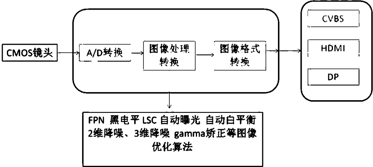
[0025] An image processing method for an ultra-fine flexible electronic endoscope, comprising an image processing chip, the image processing chip is composed of an optical imaging module, an AD conversion module, an image processing conversion module and an image format conversion module, and the optical imaging module is a CMOS The lens, the image processing conversion module is an image reverse encoding AD processing module, and the image format conversion module is a real-time image processing system; including the following steps:

[0026] A), the external cold light source guides the light to the body through the optical fiber to irradiate the visceral mucosal surface;

[0027] B) The micro-optical lens at the front of the CMOS lens gathers the light reflected by the mucous membrane and projects it onto the photosensitive surf...


PUM


Abstract

An ultra-fine flexible electronic endoscope image processing method includes the following operating steps: A), an external cold light source guides light into the body through an optical fiber to irradiate the mucous membrane surface of the visceral organs; B), a micro optical lens at the front end of a CMOS lens collects the light reflected by the mucous membranes, projects the light to a light-sensitive surface of a CMOS image sensor, and obtains image data through a CMOS sensor configured by a controller; C), the image data is converted into a serial analog signal data stream by a high-precision coding conversion chip of an AD conversion module; D), the coded image data is sent to an image reverse coding AD processing module; E), the image reverse coding AD processing module decodes the coded analog signal and inputs the signal to a real-time image processing system for processing; and F), a video encoder performs video coding and format conversion, and outputs the signal to a display to ensure that a clear and stable endoscopic image is displayed. According to the scheme of the invention, the long-distance high-fidelity transmission of the image data can be realized, and the image quality can be greatly improved.

Description

technical field [0001] The invention belongs to the technical field of medical equipment, in particular to an image processing method of an ultrafine flexible electronic endoscope. Background technique [0002] The development of endoscopes has a history of more than 200 years. It has experienced the development stages of rigid endoscopes, fiber optic endoscopes, electronic endoscopes, and capsule endoscopes. , Industrial precision non-destructive testing, fine trace identification of the public security system and other fields have broad application prospects. my country's "National Key R&D Plan: Basic Material Technology Improvement and Industrialization Key Special Project Application Guidelines for 2016" is included in the ultra-fine diameter endoscope The direction of development will play an important role in the next ten years. [0003] Cystoscopy is one of the most basic inspection methods in urology. In the past, rigid cystoscopes made of metal were used, and the dia...

Claims


Application Information

Patent Timeline
Patent Type & Authority Applications(China)
IPC IPC(8): H04N5/225H04N5/374H04N5/378H04N5/357H04N7/18H04N7/01
CPCH04N7/01H04N7/18H04N23/555H04N23/56H04N23/54H04N23/55H04N25/60H04N25/76H04N25/75
Inventor 彭剑
Owner 上海瑞烁信息科技有限公司
Why Eureka
Try Eureka