Dilator and surgical instrument

A technology of surgical instruments and dilators, which is applied in the field of medical devices, can solve the problems of low convenience and accuracy of dilators, and achieve the effects of increasing convenience and flexibility, improving convenience and accuracy, and improving convenience

Pending Publication Date: 2018-09-18
THE FIRST AFFILIATED HOSPITAL ZHEJIANG UNIV COLLEGE OF MEDICINE
View PDF0 Cites 2 Cited by
  • Summary
  • Abstract
  • Description
  • Claims
  • Application Information

AI Technical Summary

Problems solved by technology

[0005] The purpose of the present invention is to provide a dilator to solve the technical problem of low convenience and accuracy of the dilator in the prior art

Method used


Image

  • Dilator and surgical instrument
  • Dilator and surgical instrument
  • Dilator and surgical instrument

Examples


Embodiment 1

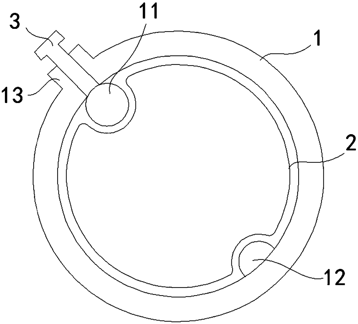
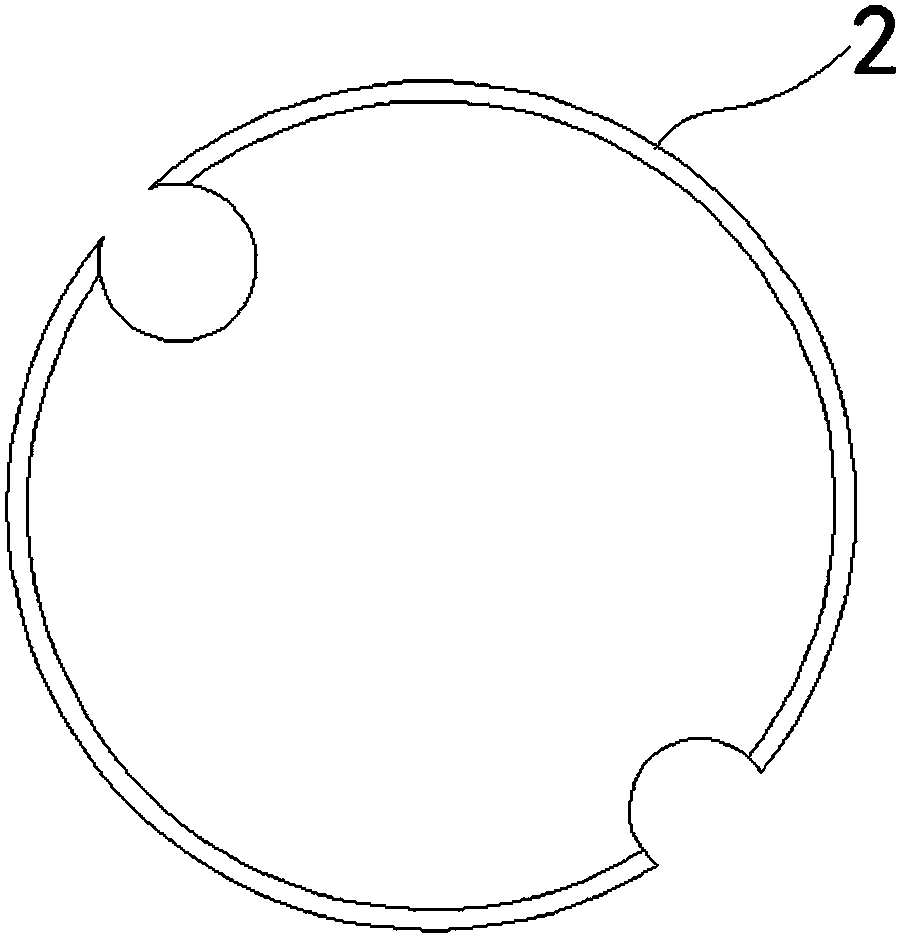
[0036] The dilator provided in this embodiment, such as Figure 1 to Figure 4 As shown, it includes an inner tube 2 and an outer tube 1; one end of the inner tube 2 is tapered; the outer tube 1 is sleeved on the inner tube 2, and the inner wall of the outer tube 1 has an endoscopic lens channel 11 and / or suction Channel 12; the endoscope lens channel 11 is used to fix the endoscope lens, and the endoscope lens is used to connect with the endoscope device; the suction channel 12 is used to connect with the aspirator.

[0037] Wherein, the inner wall of the outer tube 1 may only have the endoscopic lens channel 11 , may only have the suction channel 12 , or may have both the endoscopic lens channel 11 and the suction channel 12 .

[0038] Wherein, when there is an endoscopic lens channel 11 on the inner side wall of the outer tube 1, during operation, the inner tube 2 and the outer tube 1 are simultaneously punctured to a suitable puncture site by using the tapered end of the in...

Embodiment 2

[0059] The surgical instrument provided in this embodiment includes the dilator described in Embodiment 1. During operation, the dilator uses the end of the inner tube 2 to simultaneously puncture the inner tube 2 and the outer tube 1 to a suitable puncture site. In the tissue, the endoscopic lens can be used to observe whether the dilator reaches the target puncture site, thereby improving the accuracy of puncture. After the dilator reaches the target puncture site, the inner tube 2 is drawn out of the patient's body, and the operation can be performed. During the operation, the direction and depth of the dilator and the endoscopic lens need to be changed at any time according to the location and shape of the lesion in the brain. The dilator and the endoscopic lens can move synchronously, flexibly change the operation direction and depth, and shorten the operation time. At the same time, the suction channel 12 on the outer tube can absorb the blood and exudate in the operati...


PUM


Abstract

The invention provides a dilator and a surgical instrument, and relates to the technical field of medical instruments. The dilator comprises an inner tube and an outer tube; one end of the inner tubeis tapered; the outer tube sleeves the inner tube, and the inner side wall of the outer tube is provided with an endoscope head channel and/or a suction channel; the endoscope head channel is used forfixing an endoscope head, and the endoscope head is used for being connected with an endoscope device; the suction channel is used for being connected with an aspirator. The endoscope head can be inserted into brain tissues synchronously with the outer tube, the endoscope head can be used for observing whether or not the dilator reaches a target puncture site, during the operation, the directionand depth of the dilator are changed according to the positions and shapes of lesions in the brain, the direction and depth of a surgical operation are flexibly changed, and the operation time is shortened. The suction channel can absorb blood and seepage of a surgical view field at any time, which is beneficial for seeing the surgical view field and stopping bleeding; suction devices necessary for an operation inside dilator lumen are not needed, and the convenience and flexibility of the operation inside the dilator lumen are improved.

Description

technical field [0001] The invention relates to the technical field of medical instruments, in particular to a dilator and a surgical instrument with the dilator. Background technique [0002] The brain is the most advanced part of the nervous system. It is composed of the left and right cerebral hemispheres, and the two hemispheres are connected by horizontal nerve fibers. Cerebral hematoma and brain tumor are common diseases of the brain. Cerebral hematoma is due to trauma or cerebrovascular disease. When the blood vessels in the brain or between the brain tissue and the skull rupture, blood accumulates in the brain or between the brain and the skull. The brain tumors refer to nervous system tumors that occur in the cranial cavity, including tumors originating from the neuroepithelium, cranial nerves, meninges and germ cells, tumors of lymphoid and hematopoietic tissues, sella turcica Craniopharyngioma and pituitary tumors and metastatic tumors in the region. [0003] Wi...

Claims


Application Information

Patent Timeline
Patent Type & Authority Applications(China)
IPC IPC(8): A61B17/00A61M1/00A61B17/34
CPCA61M1/0023A61B17/00234A61B17/3415A61B2017/0034A61M1/84
Inventor 马跃辉杜朝亮
Owner THE FIRST AFFILIATED HOSPITAL ZHEJIANG UNIV COLLEGE OF MEDICINE
Why Eureka
Try Eureka