
A Segmentation Method of Epithelial Tissue in Esophagus Pathological Image

A technology for pathological images and epithelial tissue, applied in image analysis, image enhancement, image data processing, etc., to achieve the advantages of segmentation accuracy, high precision and recall rate, and less uneven staining

Active Publication Date: 2022-01-21
SHANDONG COMP SCI CENTNAT SUPERCOMP CENT IN JINAN
View PDF6 Cites 0 Cited by
  • Summary
  • Abstract
  • Description
  • Claims
  • Application Information

AI Technical Summary

Problems solved by technology

For a complete full-scan section of esophageal pathological tissue, its size is about 100,000×700,000 pixels, and it needs to occupy 1.5G of hard disk space to store on the computer. This high-resolution, large-scale image is very important for computer hardware and image analysis. Algorithms are very challenging

Method used


Image

  • A Segmentation Method of Epithelial Tissue in Esophagus Pathological Image
  • A Segmentation Method of Epithelial Tissue in Esophagus Pathological Image
  • A Segmentation Method of Epithelial Tissue in Esophagus Pathological Image

Examples


Embodiment Construction

[0042] In order to make the purpose, technical solution and advantages of the present invention clearer, the following examples are given to further describe the present invention in detail.

[0043] The implementation process of automatic segmentation of esophageal epithelial tissue in this embodiment is as follows:

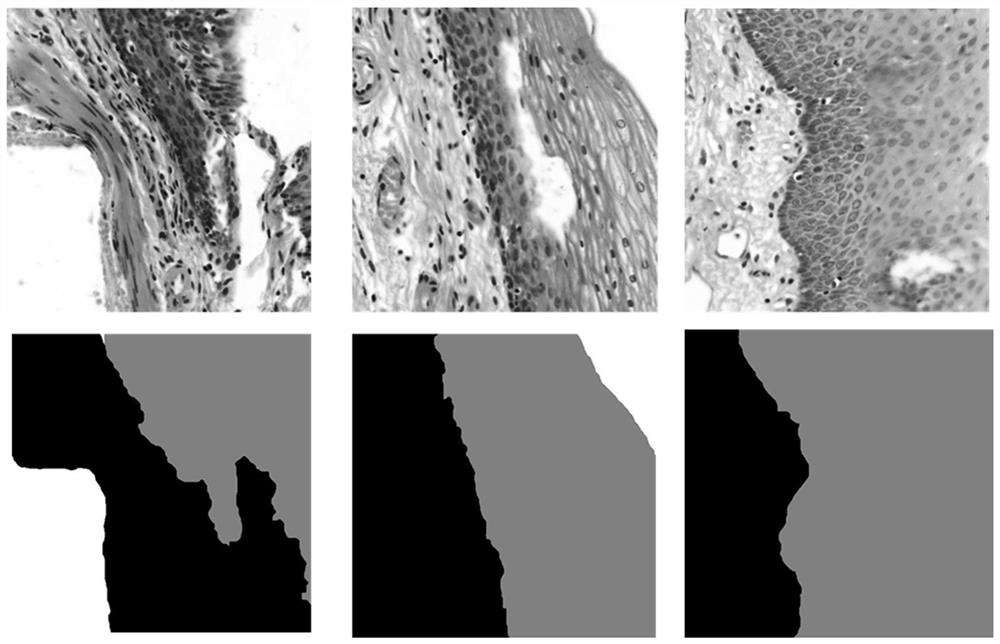
[0044] In step a), 24 H&E stained (hematoxylin-eosin stained) esophageal pathological original images (each with a size of about 1.5G) of different people are subjected to staining correction processing, such as figure 2 As shown, the images of some regions of the H&E-stained esophageal pathological section images in the present invention are given. With stain correction, slice images can be reconstructed individually according to the color of the stain, thereby facilitating quantitative analysis of slice images. Process the histopathological images of the two stains, namely Haematoxylin (H) and Eosin (Eosin, E), according to the optical density matrix, correc...


PUM


Abstract

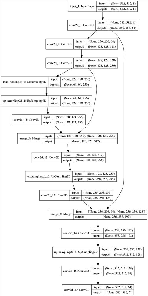
The segmentation method of epithelial tissue in the esophagus pathological image of the present invention comprises: a). Staining correction and grayscale processing; b). Selecting training and testing samples; c). Image segmentation and labeling; d). Constructing a convolutional neural network Model; e). Processing of test images; f). Acquisition of warm-up images; g). Processing of predicted heatmaps; h). Calculation of precision and recall. The epithelial segmentation method proposed in this patent is a classification at the pixel level, especially for the segmentation of the epithelial boundary area, which has obvious advantages in segmentation accuracy, and this method automatically learns effective features and expressions, avoiding complex manual features The selection process can meet the actual application requirements. The images obtained from different hospitals using different scanners can segment epithelial tissue with high precision, which is an indispensable step in image processing in the construction of computer-aided diagnosis of esophageal cancer.

Description

technical field [0001] The present invention relates to a method for segmenting epithelial tissue in pathological images of the esophagus, and more specifically, to a method for segmenting epithelial tissue in pathological images of the esophagus using a convolutional neural network model constructed from sample data. Background technique [0002] After examining a patient's biological tissue samples, the pathologist's report is often the gold standard for many diseases. With cancer in particular, a pathologist's diagnosis can have a profound impact on a patient's treatment. Pathology slide review is a very complex task that requires years of training to do well, as well as extensive expertise and experience. [0003] Esophageal cancer is a common malignant tumor in life, which seriously affects human health. The incidence of esophageal cancer in my country ranks among the highest in the world, and there are a large number of new cases of esophageal cancer every year. At ...

Claims


Application Information

Patent Timeline
Patent Type & Authority Patents(China)
IPC IPC(8): G06T7/00G06T7/11G06N3/04
CPCG06T7/0012G06T7/11G06T2207/30096G06T2207/30021G06N3/045
Inventor 牛春阳孙占全赵志刚葛菁谢迎
Owner SHANDONG COMP SCI CENTNAT SUPERCOMP CENT IN JINAN
Learn More