
A cell capture device suitable for medical testing

A capture device and cell technology, which is applied in the field of cell capture devices in medical testing, can solve problems such as single cells are not easy to separate out, and achieve the effects of avoiding leakage, ensuring air tightness, and improving precipitation efficiency

Active Publication Date: 2019-09-13
乐杰生物科技有限公司
View PDF4 Cites 0 Cited by
  • Summary
  • Abstract
  • Description
  • Claims
  • Application Information

AI Technical Summary

Problems solved by technology

[0005] The purpose of the present invention is to overcome the problem of difficult precipitation of single cells caused by the lack of precipitation structure in the prior art and the problem of concentrated force on cells caused by unreasonable design of the capture mechanism, so as to realize a kind of easy precipitation of captured cells, which is convenient for inspection personnel to use and flow The microfluidic cell capture device with no concentrated force in the channel can avoid cell lysis and cell damage caused by unreasonable capture structure to the greatest extent

Method used


Image

  • A cell capture device suitable for medical testing
  • A cell capture device suitable for medical testing
  • A cell capture device suitable for medical testing

Examples


Embodiment Construction

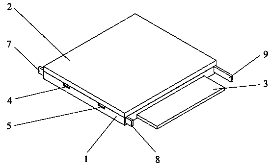
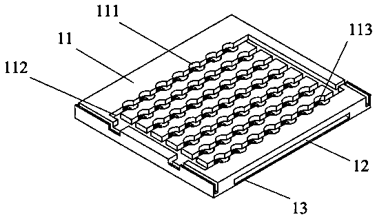
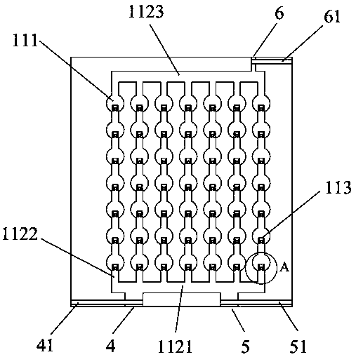
[0027] reference figure 1 , figure 2 , A cell capture device suitable for medical testing, comprising a microfluidic substrate 1, a microfluidic cover plate 2, a precipitation partition 3, a drainage inlet 4, a culture medium inlet 5, a drainage outlet 6, a first flow-stop plate 7, The second stop plate 8, the outlet opening and closing plate 9, the microfluidic substrate 1 is arranged under the microfluidic cover plate 2 and is integrated with the microfluidic cover plate 2, and the precipitation baffle 3 is movably arranged on the microfluidic substrate Inside the sheet 1, the drainage inlet 4 and the culture medium inlet 5 are opened on the front side of the microfluidic substrate 1, the drainage outlet 6 is opened on the rear side of the microfluidic substrate 1, and the first stop The flow plate 7 is movably arranged in the drainage port 4, the second flow stopper 8 is movably arranged in the culture medium inlet 5, and the outlet opening and closing plate 9 is movably arr...


PUM


Abstract

The invention relates to the technical field of cell detection, in particular to a cell capturing device for medical inspection. The cell capturing device aims at solving the problems of the prior artthat the single cells are difficult to separate out due to lack of separation structure, and the cell stress is concentrated due to unreasonable design of the capturing mechanism. The cell capturingdevice for the medical inspection comprises a microfluid substrate, a microfluid cover plate, a separation clapboard, a fluid guide inlet, a culture medium inlet, a fluid guide outlet, a first fluid blocking plate, a second fluid blocking plate, and an outlet opening and closing plate, wherein the microfluid substrate is arranged under the microfluid cover plate and is integrally bonded with the microfluid cover plate; the separation clapboard is movably arranged in the microfluid substrate. A microfluidics cell capturing device is adopted, and has the advantages that the convenience in use isrealized for an inspector; the stress concentration is avoided in the fluid channel; the cell dissolving and cell injury due to unreasonableness of capturing structure are furthest avoided.

Description

Technical field [0001] The present invention relates to the technical field of cell detection, in particular to a cell capture device suitable for medical testing. Background technique [0002] As the number one killer threatening human life and health, people are paying more and more attention to cancer research, and the metastasis of cancer cells has been plagued by scientific research and medical workers. How to quickly and effectively separate cancer cells from tissue fluid and peripheral blood Cancer cells are the primary task for further study of cancer cell metastasis and pathogenesis. At this stage, the industry mainly uses flow analyzers as the main method of cell separation. In practical applications, it has been found that the flow analyzer has problems such as complicated operation procedures, separation process affecting cell viability, and difficulty in obtaining single cells. Therefore, with the in-depth research on microelectromechanical systems in academia, the ...

Claims


Application Information

Patent Timeline
Patent Type & Authority Patents(China)
IPC IPC(8): C12M1/00
Inventor 程乐杰郝建峰李劲松侯滨蔡宏伟
Owner 乐杰生物科技有限公司
Learn More