Method for in-situ biosynthesis of multifunctional metal nanoprobe from tumor cells

A metal nanoprobe and tumor cell technology, applied in the field of material preparation, can solve the problems of toxicity and cell death, and achieve the effect of good biocompatibility

Inactive Publication Date: 2018-12-07
SOUTHEAST UNIV
View PDF15 Cites 6 Cited by
  • Summary
  • Abstract
  • Description
  • Claims
  • Application Information

AI Technical Summary

Problems solved by technology

However, these methods have more or less disadvantages. For example, the surface of metal nanoprobes synthesized based on the "one-pot cooking" method contains a large number of encapsulating agents, and most of the encapsulating agents contain some toxicity, which may cause cell damage during the process of biomarking or imaging. die

Method used


Image

  • Method for in-situ biosynthesis of multifunctional metal nanoprobe from tumor cells
  • Method for in-situ biosynthesis of multifunctional metal nanoprobe from tumor cells

Examples


Embodiment 1

[0025] A method for biosynthesizing multifunctional metal nanoprobes in situ with tumor cells, the specific steps are as follows: place HepG2 cell lines in DMEM medium containing 10% fetal bovine serum (streptomycin 100 gg / mL, penicillin 100 IU / mL) in a culture flask at 37°C in an incubator containing 5% CO2 and 95% humidity. After the cells adhere to the wall, add chloroauric acid solution (10-20 μM in the culture flask) to the culture flask, and after 12 hours of incubation, continue to add a certain concentration (for example, 10 μM) of DNA strands to the culture flask, After incubation for 12 hours, the incubated HepG2 cells were extracted by centrifugation at a rate of 2000 r / min, and the incubated HepG2 cells were washed with 10 mM barbital sodium-hydrochloric acid buffer solution for 3-5 times, and the washed cells were crushed. The incubated HepG2 cells are separated and extracted from the multifunctional gold nano-probes in the incubated HepG2 cells.
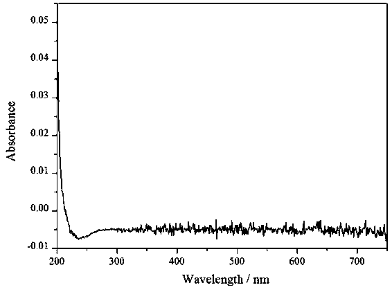
[0026] figur...

Embodiment 2

[0028] A method for biosynthesizing multifunctional metal nanoprobes in situ with tumor cells, the specific steps are as follows: Place liver cancer (HepG2) cell lines in DMEM medium containing 10% fetal bovine serum (streptomycin 100 gg / mL, penicillin 100 IU / mL) in a culture flask at 37°C in an incubator containing 5% CO2 and 95% humidity. After the cells adhere to the wall, add silver nitrate solution (10-20 μM in the culture flask) to the culture flask, and after 12 hours of incubation, continue to add a certain concentration (for example, 10 μM) of DNA strands to the culture flask, and incubate After 12 hours, the incubated HepG2 cells were extracted by centrifugation at a rate of 2000 r / min, and the incubated HepG2 cells were washed 3-5 times with 10 mM phosphate (PBS) buffer solution. the HepG2 cells, and separate and extract the multifunctional silver nanoprobes in the incubated HepG2 cells.

Embodiment 3

[0030] A method for biosynthesizing multifunctional metal nanoprobes in situ with tumor cells, the specific steps are as follows: place HepG2 cell lines in DMEM medium containing 10% fetal bovine serum (streptomycin 100 gg / mL, penicillin 100 IU / mL) in a culture flask at 37°C with 5% CO 2 , cultured in an incubator with 95% humidity. After the cells adhere to the wall, add zinc gluconate solution (concentration in the culture flask is 10-20 μM) to the culture flask, and after incubation for 12 hours, continue to add a certain concentration (for example, 10 μM) of DNA strands to the culture flask, After incubation for 12 hours, extract the incubated HepG2 cells by centrifugation at a rate of 2000 r / min, wash the incubated HepG2 cells with 10 mM Tris-hydrochloric acid buffer solution for 3-5 times, break and wash the incubated HepG2 cells cells, separating and extracting the multifunctional zinc nano-probes in the incubated HepG2 cells.


PUM

PropertyMeasurementUnit
particle diameteraaaaaaaaaa

Abstract

The invention relates to a method for in-situ biosynthesis of a metal nanoprobe from tumor cells. The method comprises the following steps: subjecting a metal soluble salt, DNA or RNA strands and tumor cells to co-incubation in a cell culture medium for 12-24 hours, then centrifugally extracting incubated tumor cells at a rate to 1000-2500 r/min; cleaning the incubated tumor cells with a buffer solution 3-5 times; breaking the cleaned incubated tumor cells; and separating and extracting the multifunctional metal nanoprobe in the incubated tumor cells. The multifunctional metal nanoprobe of theinvention has good biocompatibility and is greatly enhanced in application potential in fields of biomarkers, imaging, sensing, tumor therapy and the like.

Description

technical field [0001] The invention relates to the field of material preparation, in particular to a method for biosynthesizing multifunctional metal nanoprobes in situ with tumor cells. Background technique [0002] In situ fluorescence bioimaging is of great significance in the diagnosis and analysis of tumors. Generally, the target molecules, tumor cells or tissues are marked and combined with optical microscopic imaging technology to realize the visual analysis and monitoring of the biological process of tumor cells or tissues and the process of therapeutic drugs on tumor cells or tissues. In recent years, due to the intense research on metal nanoprobes, metal nanoprobes have become emerging materials for labeling tumor cells or tissues. [0003] In recent years, metal nanoprobes (such as gold, silver, etc.) with good biocompatibility and high fluorescence intensity have been favored by researchers. Their size is usually less than 2 nm, and they have strong visible-nea...

Claims


Application Information

Patent Timeline
Patent Type & Authority Applications(China)
IPC IPC(8): G01N21/64
CPCG01N21/6486G01N2021/6497
Inventor 王雪梅姜晖李春梅阮俊陈芸张航
Owner SOUTHEAST UNIV
Why Eureka
Try Eureka