Cell preparation reagent, kit and usage
A cell preparation and kit technology, applied in the field of cell preparation reagents, can solve problems such as pollution, increase products, etc., and achieve the effects of convenient operation, improved adhesion, and improved processing quality.
- Summary
- Abstract
- Description
- Claims
- Application Information
AI Technical Summary
Problems solved by technology
Method used
Image
Examples
Embodiment 1
[0042]
[0043]
[0044] Prepare 100g of sample processing reagent according to the above components, dissolve the above components in ultrapure water, stir and mix, and make up 100g of ultrapure water.
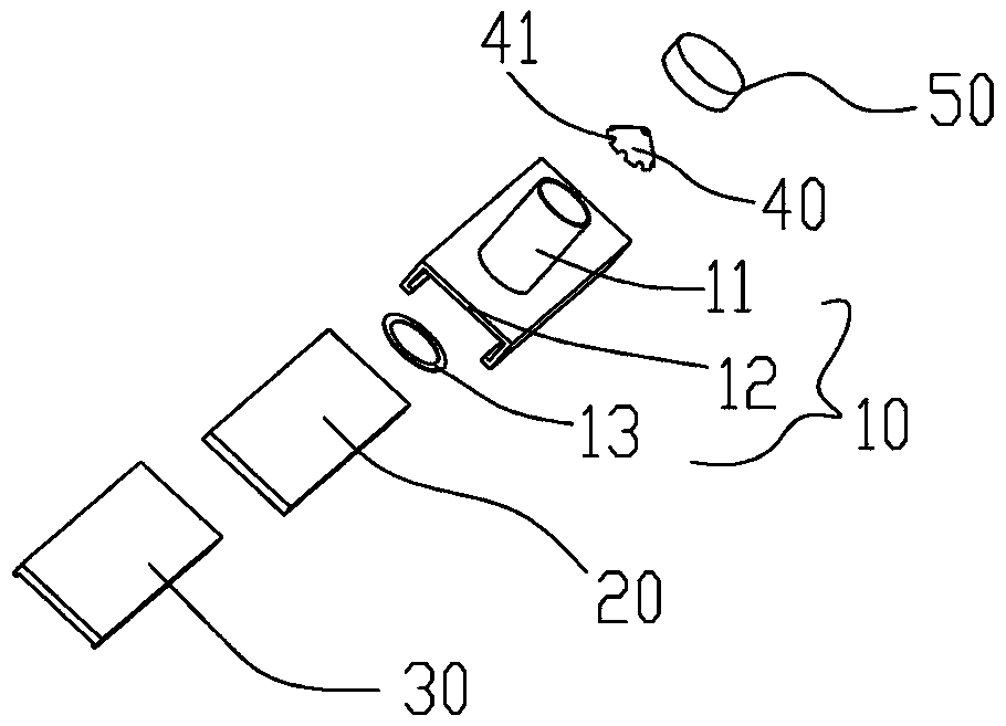
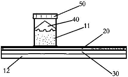
[0045] Packing process: Take a 10ml quantitative dispenser, quantitatively dispense 2ml of the sample processing reagent into the tube body of the cell production kit, and seal it with a sealed cover. 5 pieces are used as a group, and they are plastic-sealed in a blister box. into merchandise sales.
[0046] Instructions:
[0047] Step 1: Remove the sealing cap, add the sample, and re-fix the sealing cap on the reagent storage tube;
[0048] Step 2: Place the kit on the cell smear centrifuge;
[0049] Step 3: centrifuge at 1500 rpm for 2 minutes, discard the supernatant;
[0050] Step 4: Take the slides and fix them in 95% ethanol.
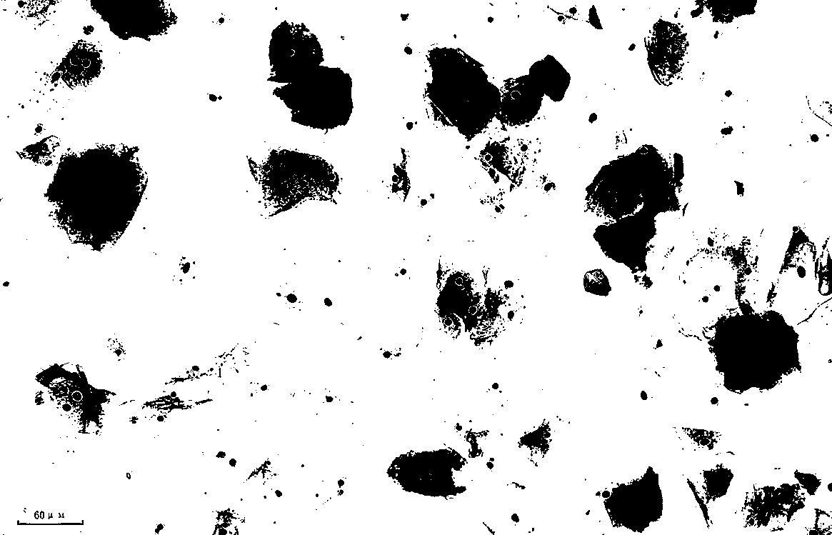
[0051] According to the above-mentioned steps, the film production is completed, and Papanicolaou staining is performed, and the effec...
Embodiment 2
[0053]
[0054]
[0055] Prepare 100g of sample processing reagent according to the above components, dissolve the above components in ultrapure water, stir and mix, and make up 100g of ultrapure water.
[0056] Packing process: Take a 10ml quantitative dispenser, quantitatively dispense 2ml of the sample processing reagent into the tube body of the cell production kit, and seal it with a sealed cover. 10 pieces are used as a group, and they are plastic-sealed in a blister box. into merchandise sales.
[0057] Instructions:
[0058]Step 1: Remove the sealing cap, add the sample, and re-fix the sealing cap on the reagent storage tube;
[0059] Step 2: Place the kit on the cell smear centrifuge;
[0060] Step 3: centrifuge at 1100 rpm for 2 minutes, discard the supernatant;
[0061] Step 4: Take the slides and fix them in 95% ethanol.
[0062] According to the above-mentioned steps, the film production is completed, and Papanicolaou staining is performed, and the effec...
Embodiment 3
Make up 100g
[0065] Prepare 100g of sample processing reagent according to the above components, dissolve the above components in ultrapure water, stir and mix, and make up 100g of ultrapure water.
[0066] Packing process: Take a 10ml quantitative liquid dispenser, quantitatively dispense 2ml of sample processing reagent into the tube body of the tablet maker, and seal it with a sealing cover. 25 pieces are used as a group, and plastic-sealed in a blister box to make a product Sales.
[0067] Instructions:
[0068] Step 1: Remove the sealing cap, add the sample, and re-fix the sealing cap on the reagent storage tube;
[0069] Step 2: Place the kit on the cell smear centrifuge;
[0070] Step 3: Spin up the centrifuge at 1100 speed for 3 minutes, discard the supernatant;
[0071] Step 4: Take the slides and fix them in 95% ethanol.
[0072] According to the above-mentioned steps, the film production is completed, and Papanicolaou staining is performed, and the effe...
PUM
 Abstract
Description
Claims
Application Information
 - R&D
- Intellectual Property
- Life Sciences
- Materials
- Tech Scout
- Unparalleled Data Quality
- Higher Quality Content
- 60% Fewer Hallucinations
Browse by: Latest US Patents, China's latest patents, Technical Efficacy Thesaurus, Application Domain, Technology Topic, Popular Technical Reports.
© 2025 PatSnap. All rights reserved.Legal|Privacy policy|Modern Slavery Act Transparency Statement|Sitemap|About US| Contact US: help@patsnap.com


