Isolated culture method for human endometrial tissue-derived endothelial progenitor cell

A technology of endothelial progenitor cells and endometrium, applied in the field of endothelial progenitor cells with strong angiogenesis ability, can solve the problems of limited angiogenesis ability and limited source of endothelial progenitor cells, and achieve the effect of strong angiogenesis ability

Active Publication Date: 2018-12-21
SHANXI MEDICAL UNIV
View PDF1 Cites 2 Cited by
  • Summary
  • Abstract
  • Description
  • Claims
  • Application Information

AI Technical Summary

Problems solved by technology

However, problems such as the limited source of existing endothelial progenitor cells and limited angiogenesis ability still exist.

Method used


Image

  • Isolated culture method for human endometrial tissue-derived endothelial progenitor cell
  • Isolated culture method for human endometrial tissue-derived endothelial progenitor cell
  • Isolated culture method for human endometrial tissue-derived endothelial progenitor cell

Examples


Embodiment 1

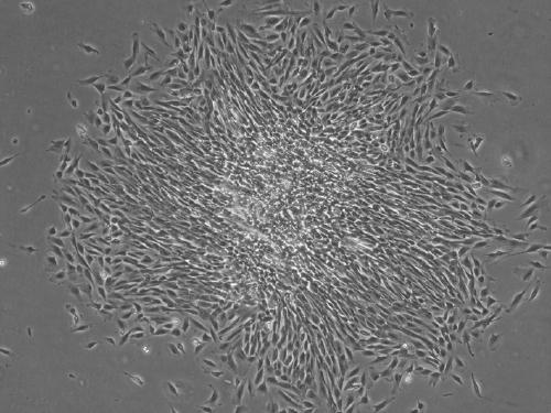
[0040] Example 1: Isolation and culture of human endometrium endothelial progenitor cells.

[0041] 1) Obtain human endometrial tissue, place it in sterile 1×PBS, and move it into the ultra-clean operating table as soon as possible.

[0042] 2) In a sterile culture vessel, the human endometrial tissue was repeatedly washed with PBS 3 times to remove surface blood and other impurities.

[0043] 3) Fully mince the cleaned human endometrial tissue to less than 1mm 3 .

[0044] 4) Add 3 times the volume of PBS digestion solution containing 300 µg / ml collagenase III and 40 µg / ml DNase I to the minced human endometrial tissue, shake and digest in a 37°C water bath at a shaking speed of 110rpm for 45min, and extract the digested product The supernatant was neutralized by adding 10% FBS-DMEM / F12 at a volume ratio of 1:1 to terminate the digestion.

[0045] 5) Add the above digestion solution to the undigested tissue fragments again, and repeat the digestion process of step 4).

[...

Embodiment 2

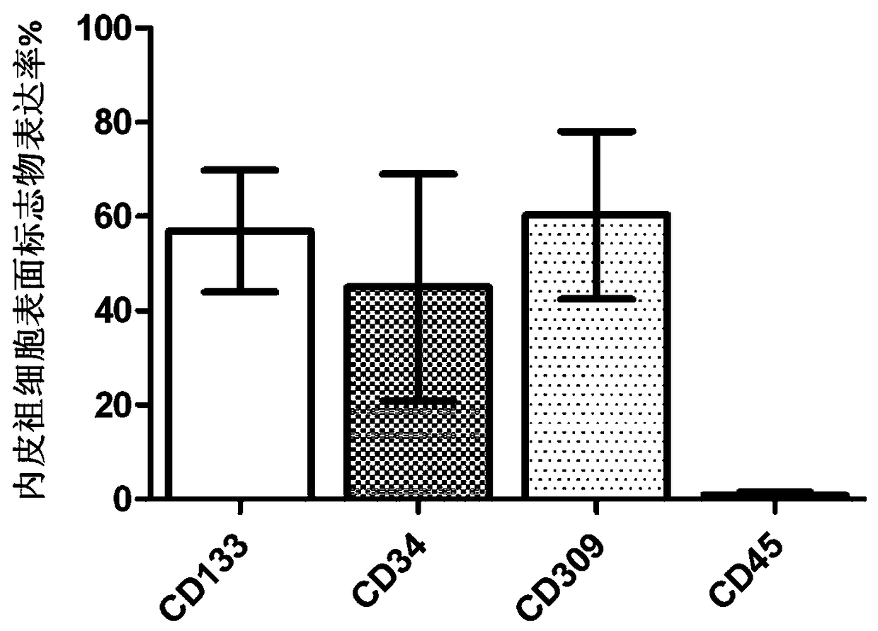
[0060] Example 2: Flow cytometric identification of human endometrial endothelial progenitor cells.

[0061] 1) The surface markers of human endometrium endothelial progenitor cells are CD133, CD34, CD309 and CD45.

[0062] 2) Select the endothelial progenitor cells cultured to the 7th day, 14th day, and 21st day respectively, and after routine digestion, stain with the antibody corresponding to the above-mentioned surface markers.

[0063] 3) Detection of surface markers of human endometrium endothelial progenitor cells by flow cytometry.

[0064] figure 2 The results of flow cytometry identification on the 14th day of culture are given, and the histogram shows the positive percentage of various surface markers.

[0065] figure 2 showed that the isolated and cultured human endometrial endothelial progenitor cells displayed CD133 + , CD34 + , CD309 + and CD45 - The characteristics of the surface markers are consistent with the characteristics of the surface markers of...

Embodiment 3

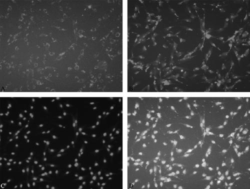
[0066] Example 3: Functional identification of human endometrium endothelial progenitor cells uptake Dil-ac-LDL and bind FITC-UEA-1.

[0067] 1) Sow human endometrial endothelial progenitor cells in a 6-well plate, place glass slides in advance in the 6-well plate, add EGM-2 culture medium, observe the growth of the cells, and wait until the cells are fused to 50-70%.

[0068] 2) Add 1ml of EGM-2 containing 10µg / ml Dil-ac-LDL into the culture well, and culture in the dark for 18h in a cell culture incubator at 37°C.

[0069] 3) After 18 hours, suck out the EGM-2 solution containing Dil-ac-LDL, wash 3 times with PBS, and fix with 4% paraformaldehyde for 30 minutes at room temperature.

[0070] 4) Wash with PBS 3 times.

[0071] 5) Add 1ml of 10µg / ml FITC-UEA-1 working solution and incubate at 37°C for 2h in the dark.

[0072] 6) After 2 hours, suck out the FITC-UEA-1 working solution and wash with PBS 3 times.

[0073] 7) Add 1ml of 3µg / ml DAPI and incubate at 37°C for 10min...


PUM


Abstract

The invention relates to an isolated culture method for a human endometrial tissue-derived endothelial progenitor cell. The endometrial tissue-derived endothelial progenitor cell is isolated from thehuman endometrial tissue. After an enzymolysis method is used for digesting the human endometrial tissue to obtain single-cell suspension, a density gradient centrifugation method is adopted to isolate to obtain a cell group which contains endothelial progenitor, the cell group is inoculated in an endothelial progenitor cell culture medium to carry out culture and passage purification, and finally, a new endothelial progenitor cell with an ultrastrong angiogenesis ability is obtained. An experiment proves that the endothelial progenitor cell has self proliferation and clone sample growth abilities, has an endothelial progenitor cell surface marker, also has the abilities of uptaking Dil-ac-LDL, combining with FITC-UEA-1 and forming tubular structures in vitro, and can be used for preparingmedicines capable of preventing or treating ischemic diseases.

Description

technical field [0001] The invention relates to a new endothelial progenitor cell, especially an endothelial progenitor cell with strong angiogenesis ability extracted from human endometrium tissue. The invention also relates to a method for isolating and culturing the endometrium endothelial progenitor cells. Background technique [0002] In recent years, with the changes in people's living standards and living habits, the number of patients with various ischemic diseases including cardiovascular and cerebrovascular diseases and the resulting medical and social problems have shown a continuous growth trend, with high incidence and fatal It has become a major disease that endangers human health. [0003] The so-called ischemic disease is a kind of poor tissue perfusion due to vascular stenosis or occlusion, resulting in insufficient blood supply to ischemic local tissue cells, ischemic injury, necrosis, and damage to local and even systemic tissues and organs. For ischemic...

Claims


Application Information

Patent Timeline
Patent Type & Authority Applications(China)
IPC IPC(8): C12N5/071A61K35/36A61P9/10
CPCA61P9/10A61K35/36C12N5/0628C12N2509/00
Inventor 解军李仁科范雪梅宋慧芳何生平毅
Owner SHANXI MEDICAL UNIV
Why Eureka
Try Eureka