Anti-cancer drug assessment method
An evaluation method and technology of anti-cancer agents, which are applied in the fields of biochemical equipment and methods, tumor/cancer cells, compound screening, etc., to achieve the effect of simple implementation
- Summary
- Abstract
- Description
- Claims
- Application Information
AI Technical Summary
Problems solved by technology
Method used
Image
Examples
Embodiment 1
[0137] [Example 1] Fabrication of a cell structure having a vascular structure
[0138] Fabricate a cell structure composed of fibroblasts and vascular endothelial cells and have a vascular network structure, and observe the vascular network structure.
[0139] As the cell structure including the vascular network structure, human neonatal skin fibroblasts (manufactured by Lonza, CC-2509, Normal Human Dermal Fibroblasts: NHDF), human umbilical cord vein endothelial cells (manufactured by Lonza, CC-2509) were used. 2517A, Human Umbilical Vein Endothelial Cell: HUVEC) A cell structure formed by two types of cells. In addition, as a cell culture container, a Transwell insert cell culture device (manufactured by Corning, product number: #3470) was used, and as a medium, a 10% by volume bovine serum (manufactured by System Biosciences, EXO-FBS-50A-1) was used. D-MEM (manufactured by Wako Pure Chemical Industries, Ltd., product number: 043-30085) and 1% by volume of penicillin / strep...
Embodiment 2
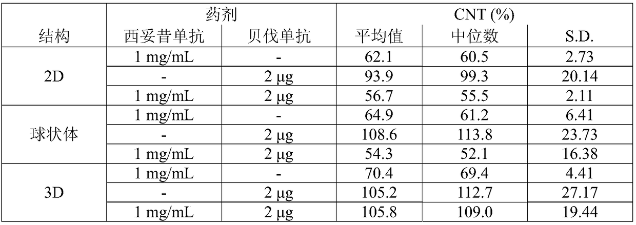
[0148] The anticancer effect of the anticancer agent doxorubicin was evaluated using a cell structure composed of fibroblasts, vascular endothelial cells, and cancer cells and having a vascular network structure.
[0149] As a cell structure containing cancer cells and a vascular network structure, human neonatal skin fibroblasts (Normal Human Dermal Fibroblasts: NHDF) (Lonza, CC-2509) and human umbilical cord vein endothelial cells (Lonza , CC-2517A, HUVEC) (Lonza Corporation, CC-2517), the top surface of the cell structure formed by the two types of cells formed by the human colorectal adenocarcinoma cell line HT29 (ATCC code: HTB-38TM) Cell structure of cancer cell layer. In addition, as a cell culture vessel, a Transwell insert cell culture device (manufactured by Corning, product number: #3470) was used, and as a medium, a 10% by volume bovine serum (manufactured by Corning, product number: #35-010- CV) and D-MEM (manufactured by Wako Pure Chemical Industries, Ltd., prod...
Embodiment 3
[0169] The anticancer effect of the anticancer agent doxorubicin was evaluated using a cell structure in which the vascular network structure was formed in layers and a cell structure in which the vascular network structure was similarly dispersed throughout the structure.
[0170] The cell culture vessel, medium, and doxorubicin were the same as those used in Example 2.
[0171]
[0172] Will 2×10 6 NHDF and 3×10 4 A cell structure was constructed in the same manner as in Example 2, except that each HUVEC was suspended in Tris-HCl buffer containing heparin and collagen. The obtained cell structure is a cell structure in which a cancer cell layer is laminated on a layer in which the vascular network structure is dispersed throughout the structure (mixed layer (21 layers) of NHDF (20 layers) and HUVEC (1 layer)-HT29 (1 story)).
[0173]
[0174] First, the 1×10 6 Each NHDF was suspended in a Tris-HCl buffer containing heparin and collagen (0.1 mg / mL heparin, 0.1 mg / mL c...
PUM
 Abstract
Description
Claims
Application Information
 - R&D
- Intellectual Property
- Life Sciences
- Materials
- Tech Scout
- Unparalleled Data Quality
- Higher Quality Content
- 60% Fewer Hallucinations
Browse by: Latest US Patents, China's latest patents, Technical Efficacy Thesaurus, Application Domain, Technology Topic, Popular Technical Reports.
© 2025 PatSnap. All rights reserved.Legal|Privacy policy|Modern Slavery Act Transparency Statement|Sitemap|About US| Contact US: help@patsnap.com


