
A method and apparatus for image region segmentation of ischemic stroke

An ischemic stroke and region segmentation technology, applied in the field of image processing, can solve the problems of multiple labor costs, dispersion, and low segmentation accuracy, and achieve the effects of improving segmentation accuracy, avoiding errors, and saving labor costs.

Active Publication Date: 2019-03-01
BEIJING SENSETIME TECH DEV CO LTD
View PDF11 Cites 14 Cited by
  • Summary
  • Abstract
  • Description
  • Claims
  • Application Information

AI Technical Summary

Problems solved by technology

The whole process is relatively scattered, requiring too much labor cost, and due to the interference of too many human factors, the final segmentation accuracy is not high

Method used


Image

  • A method and apparatus for image region segmentation of ischemic stroke
  • A method and apparatus for image region segmentation of ischemic stroke
  • A method and apparatus for image region segmentation of ischemic stroke

Examples


Embodiment Construction

[0026] The embodiments of the present application will be described below in conjunction with the drawings in the embodiments of the present application.

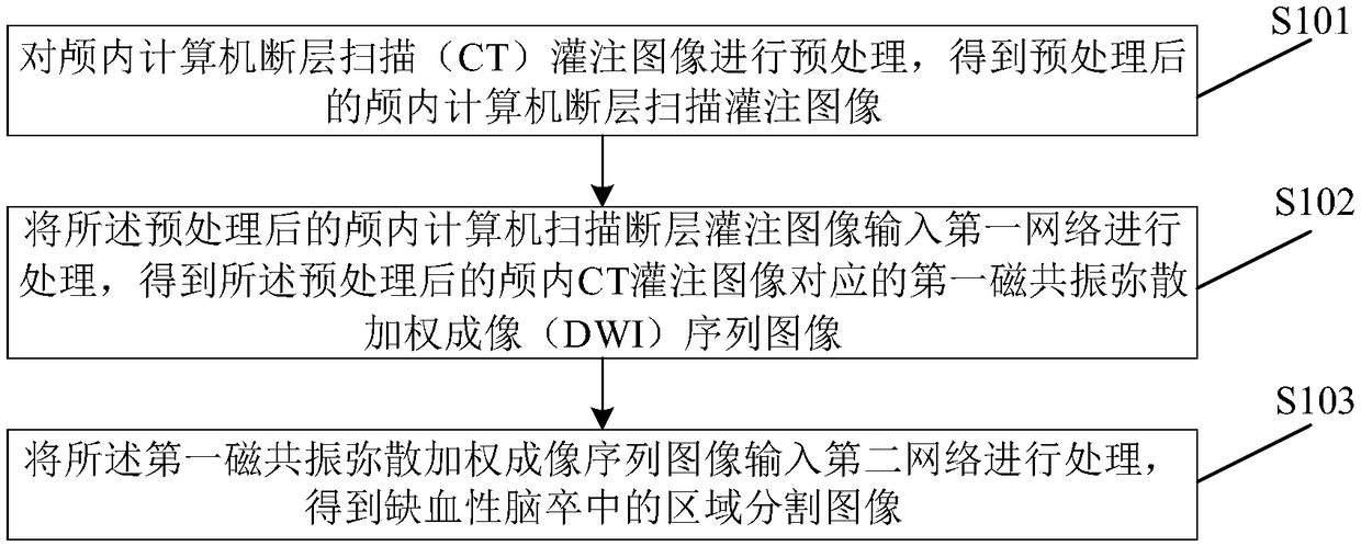
[0027] See figure 1 , figure 1 It is a schematic flowchart of a method for segmentation of an ischemic stroke image provided by an embodiment of the present application.

[0028] S101. Preprocess the intracranial computed tomography (CT) perfusion image to obtain a preprocessed intracranial computed tomography perfusion image.

[0029] Normalize the brain computer tomography image obtained by CT (Computed Tomography) scan through the Batch Norm layer without learning ability to obtain a stable mean and standard deviation, and complete the first intracranial computer tomography perfusion imaging The preprocessing of the CTP timing chart, and the CTP timing chart in the skull after preprocessing.

[0030] Performing the above normalization process when training the network can make the network have better generalization ability in th...


PUM


Abstract

A method and apparatus for image region segmentation of ischemic stroke are disclosed. The method comprises the following steps: preprocessing intracranial computed tomography (CT) perfusion images toobtain preprocessed intracranial CT perfusion images; inputting the pre-processed intracranial CT perfusion image into a first network for processing to obtain a first magnetic resonance diffusion weighted imaging (DWI) sequence image corresponding to the pre-processed intracranial CT perfusion image; the first MR diffusion-weighted imaging sequence image is input into a second network for processing to obtain a region segmentation image of ischemic stroke. A corresponding device is also disclosed. The first DWI sequence image is obtained by convolution operation of CT perfusion parameter map, and then the region segmentation image is obtained according to the first DWI sequence image. The CT perfusion image can be divided into cerebral infarction region, ischemia penumbra region and background region automatically and efficiently.

Description

Technical field [0001] This application relates to the field of image processing technology, and in particular to a method and device for segmentation of ischemic stroke image regions. Background technique [0002] Ischemic stroke is a common cerebrovascular disease, which has always seriously endangered human health, and its senile morbidity, disability and mortality rates are very high. How to diagnose ischemic stroke quickly and effectively is an important research topic in current clinical work. Early detection of ischemic stroke is very important, especially the correct detection of hyperacute phase, which can promptly guide and take measures to reduce brain cell necrosis in the ischemic penumbra area around the infarct focus. The diagnosis and treatment of sexual stroke play a major role. [0003] At present, the diagnosis of stroke ischemic penumbra and cerebral infarction is still in the manual AIF (Arterial Input Function) labeling of the CTP (Computed Tomography Perfusi...

Claims


Application Information

Patent Timeline
Patent Type & Authority Applications(China)
IPC IPC(8): G06T7/11G06T7/136
CPCG06T2207/10081G06T7/11G06T7/136
Inventor 宋涛刘蓬博
Owner BEIJING SENSETIME TECH DEV CO LTD
Features
  • Generate Ideas
  • Intellectual Property
  • Life Sciences
  • Materials
  • Tech Scout
  • Unparalleled Data Quality
  • Higher Quality Content
  • 60% Fewer Hallucinations
Learn More