Dual-mode image automatic fusion method

A fusion method and image technology, which is applied in the field of medical devices, can solve the problems of limited OCT tissue imaging depth, inability to reflect the endometrium, and limited resolution of fine structure images, achieving good fusion effects and speed improvements

Active Publication Date: 2019-04-05
INNERMEDICAL CO LTD
View PDF10 Cites 8 Cited by
  • Summary
  • Abstract
  • Description
  • Claims
  • Application Information

AI Technical Summary

Problems solved by technology

[0003]Because IVUS uses the reflection of sound waves, it is beneficial to display deep structures and is not affected by blood flow, so there is no need to block blood flow during the detection process. However, the resolution of fine structure images is limited
The strong resolution of OCT can describe the intima of the vessel

Method used


Image

  • Dual-mode image automatic fusion method
  • Dual-mode image automatic fusion method
  • Dual-mode image automatic fusion method

Examples


Embodiment Construction

[0050] The preferred embodiments of the present invention will be further described in detail below.

[0051] A dual-mode image automatic fusion method, comprising the following steps:

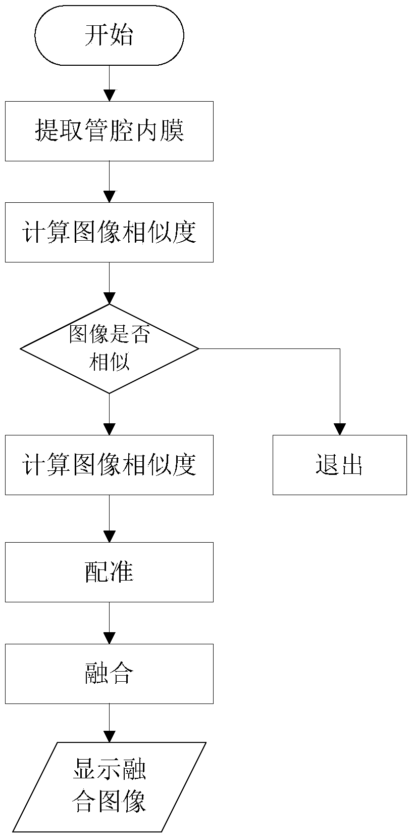
[0052] 1. Whether to perform registration, use the following steps to judge, such as figure 1 shown;

[0053] (1) Based on the snake model, extract the intima points of IVUS and OCT respectively, collect the corresponding points from the continuous points of the intima at intervals of [0-360] degrees counterclockwise, and calculate the distance from the point to the image center to obtain IVUS, OCT intima point to image center distance sequence N is the number of collection points on the intima.

[0054] (2) To calculate the similarity between two distance sequences, the Euclidean distance between the sequences, histogram matching, correlation coefficient, etc. can be used as the similarity measure of the two sequences. Among them, the Pearson (Pearson) correlation coefficient is defined as...


PUM


Abstract

The invention provides a dual-mode image automatic fusion method which comprises the following steps: respectively obtaining distance sequences from IVUS and OCT (Optical Coherence Tomography) inner membrane points to the center of an image on the basis of a sinake model; Calculating scaling coefficients of the OCT image and the IVUS image blood vessel lumen through the average inner membrane radius of the IVUS image and the average inner membrane radius of the OCT image blood vessel lumen; Rotating the IVUS image according to a preset angle range and a preset step length; calculating mutual information of the rotated IVUS image and the OCT image; when the mutual information is the maximum value, the corresponding angle serves as a reference, adopting a circumferential correlation algorithm for estimating the actual rotation angle, and after IVUS and OCT images are registered, adoptinga Poisson fusion algorithm for fusing the converted IVUS image and the converted OCT original image. According to the technical scheme, the advantages of IVUS imaging and OCT imaging are brought into full play, and the speed of the fusion process is increased.

Description

technical field [0001] The invention belongs to the technical field of medical devices, and in particular relates to a dual-mode image automatic fusion method. Background technique [0002] Intravascular Ultrasound (IVUS), as a branch of medical ultrasound, is the result of using ultrasonic principles to detect the inside of blood vessels, blood vessel walls and surrounding tissues. It is an invasive tomographic imaging technology for disease diagnosis and treatment. Optical Coherence Tomography (OCT) is currently the highest-resolution intravascular imaging technology, which can better display images of various layers of the vessel wall, and has more accurate and high-contrast imaging of plaques and intima. IVUS can obtain larger imaging depth and range based on the principle of ultrasound imaging, but its image clarity is insufficient; OCT can obtain high-definition imaging based on the principle of optical imaging, but the imaging depth and range are insufficient. The te...

Claims


Application Information

Patent Timeline
IPC IPC(8): G06T5/50G06T7/00G06T7/33
CPCG06T5/50G06T7/0012G06T7/344G06T2207/30101
Inventor 白晓淞
Owner INNERMEDICAL CO LTD
Why Eureka
Try Eureka