
A heart left ventricle segmentation method based on a deep full convolutional neural network

A convolutional neural network and left ventricle technology, applied in the field of heart left ventricle segmentation, can solve problems such as low segmentation accuracy and loss of fine object information, and achieve the effect of time-consuming and labor-intensive solutions

Active Publication Date: 2019-04-05
ZHEJIANG UNIV
View PDF3 Cites 37 Cited by
  • Summary
  • Abstract
  • Description
  • Claims
  • Application Information

AI Technical Summary

Problems solved by technology

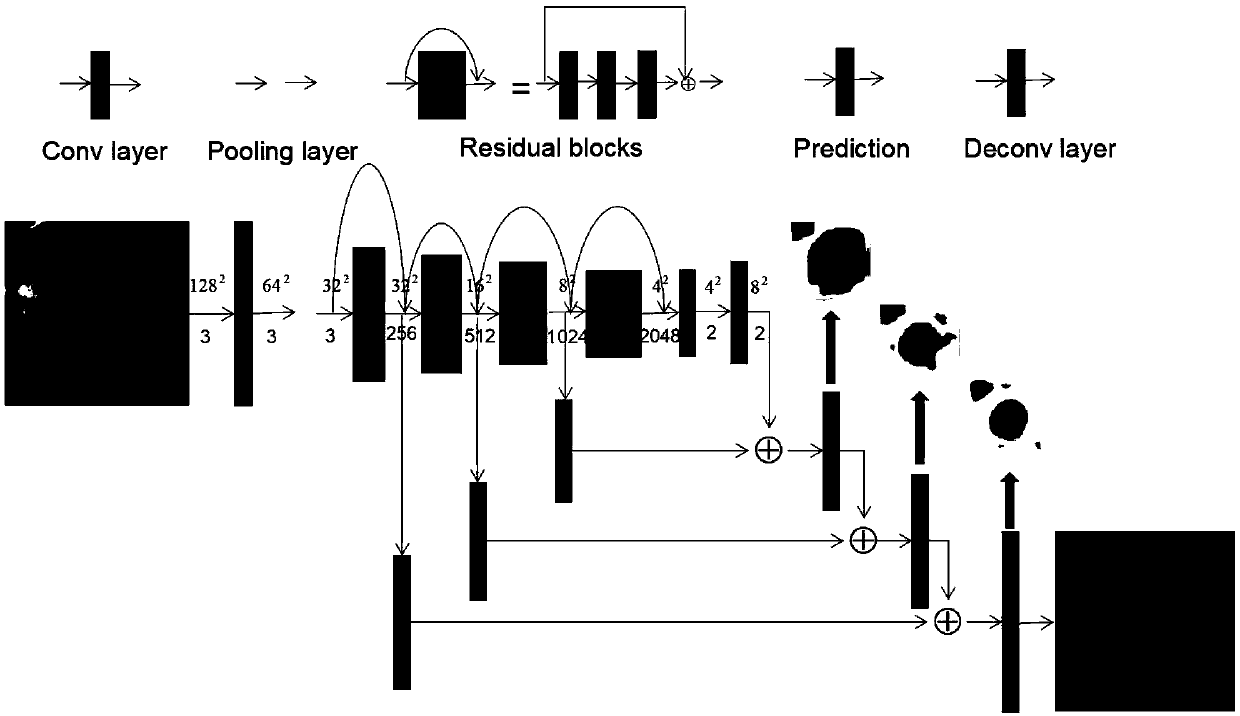
These include FCN architecture (Tran P V.A Fully Convolutional Neural Network for Cardiac Segmentation in Short-Axis MRI[J].2016.) and UNet architecture (Ronneberger O, FischerP, Brox T.U-Net: Convolutional Networks for Biomedical ImageSegmentation[J].2015 .) for left ventricle segmentation; the general idea behind FCN is to use the downsampling path to learn relevant features at various spatial scales, and then use the upsampling path to combine features predicted at the pixel level. The network does not overcome this segmentation difficulty when the size is at the apex or end-systole of the heart, because fine object information may be lost during the downscaling process of the maximization layer in the network
UNet is one of the commonly used segmentation models in medical imaging. The network obtains the segmentation map through multiple deconvolutions. In addition, the convolution layer information of the corresponding size at the front end of the network is added when the deconvolution is sampled, so that more details can be obtained. It is preserved, but the network does not pay more attention to the pixels of the segmentation boundary, and there will also be a problem of low segmentation accuracy at the apex of the heart or at the end of systole

Method used


Image

  • A heart left ventricle segmentation method based on a deep full convolutional neural network
  • A heart left ventricle segmentation method based on a deep full convolutional neural network
  • A heart left ventricle segmentation method based on a deep full convolutional neural network

Examples


Embodiment Construction

[0024] In order to describe the present invention more clearly, the technical solution of the present invention will be described in detail below in conjunction with the accompanying drawings and specific embodiments.

[0025] The present invention is based on the heart left ventricle segmentation method of deep fully convolutional neural network, and specific implementation steps are as follows:

[0026] S1. Obtain the complete cardiac magnetic resonance image of the subject, as well as the corresponding manually drawn left ventricular endocardial contour line. The subject's complete cardiac MRI short-axis images only contain ordinary cine sequences, such as figure 2 As shown, that is, the coronal, sagittal, and axial three-direction positioning maps are simultaneously made on the subject by the magnetic resonance instrument. The imaging range is from the bottom of the heart and the root of the great blood vessels to the apex of the heart. We only screen short-axis cardiac i...


PUM


Abstract

The invention discloses a heart left ventricle segmentation method based on a deep full convolutional neural network. According to the method, a deep learning idea is introduced into heart magnetic resonance short-axis image left ventricle segmentation; The process is mainly divided into a training stage and a prediction stage, in the training stage, a preprocessed 128 * 128 heart magnetic resonance image serves as input, a manually processed label serves as a label of a network to be used for calculating errors, and along with increase of training iteration times, the error of a training setand the error of a verification set are gradually reduced; And in the test stage, inputting data in the test set into the trained model, and finally outputting prediction of each pixel by the networkto generate a segmentation result. According to the method, segmentation of the heart magnetic resonance short-axis image is achieved from the perspective of data driving, the problem that manual outline drawing is time-consuming and labor-consuming is effectively solved, the defects of a traditional image segmentation algorithm can be overcome, and high-precision and high-robustness left ventricle segmentation is achieved.

Description

technical field [0001] The invention belongs to the technical field of medical image analysis, and in particular relates to a heart left ventricle segmentation method based on a deep fully convolutional neural network. Background technique [0002] In recent years, cardiovascular disease has become one of the number one killers of human life and health. With the improvement of people's living standards and the rapid development of modern medicine, early diagnosis and risk assessment of cardiovascular diseases have become important conditions for improving human living standards. In addition, with the continuous advancement of medical technology, imaging equipment capable of dynamic imaging of the heart mainly includes Magnetic Resonance Imaging (MRI), Computed Tomography (X-Ray Computer Tomography, CT) and Ultrasonic Imaging (Ultrasonic Imaging, US )Wait. Cardiac magnetic resonance imaging has good soft tissue contrast, no radioactivity, no need to inject or take tracers, a...

Claims


Application Information

Patent Timeline
IPC IPC(8): G06T7/11G06N3/04
CPCG06T7/11G06T2207/30048G06T2207/20084G06T2207/20081G06T2207/10088G06N3/045
Inventor 刘华锋陈明强
Owner ZHEJIANG UNIV
Learn More