4-axis 3D-printing tubular medical stent and preparation method thereof

A technology of 3D printing and printing tubes, applied in the field of medical stents, can solve problems such as difficulty in preparing thin-walled porous tubular stents, and achieve good application prospects, simple methods, and the effect of preventing thrombosis

Active Publication Date: 2019-05-03
DONGHUA UNIV +1
View PDF10 Cites 10 Cited by
  • Summary
  • Abstract
  • Description
  • Claims
  • Application Information

AI Technical Summary

Problems solved by technology

[0007] The technical problem to be solved by the present invention is to provide a 4-axis 3D printing tubular medical stent and its preparation method, which solves the fundamental problem that extrusion 3D printing is difficult to prepare a thin-walled porous tubular stent, and the prepared medical stent has structural advantages. Accurate and controllable multi-level pore structure, excellent elasticity and sutureability in mechanical properties, simple and fast method, suitable for a variety of biological materials, and has good application prospects

Method used


Image

  • 4-axis 3D-printing tubular medical stent and preparation method thereof
  • 4-axis 3D-printing tubular medical stent and preparation method thereof
  • 4-axis 3D-printing tubular medical stent and preparation method thereof

Examples


preparation example Construction

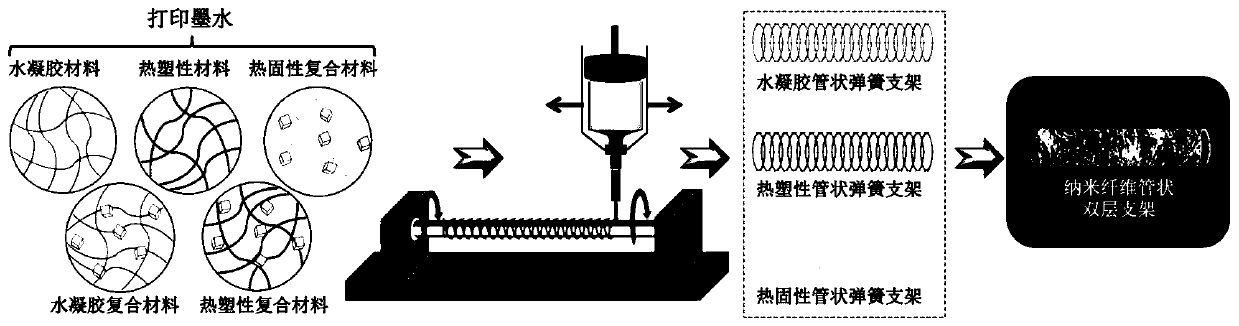
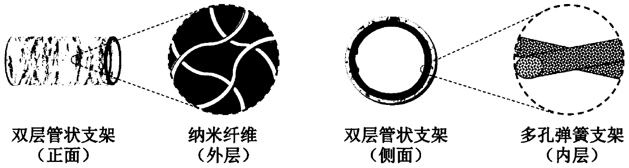
[0057] 1.4 Preparation of double-layer tubular scaffold

[0058] A gelatin / hexafluoroisopropanol solution (12%, w / v) was prepared as an electrospinning solution, drawn into a 5ml syringe, and installed on a microsyringe pump. The above-mentioned tubular PGS biological spring is threaded into a metal receiver of corresponding specifications, and installed on a rotating receiving device with adjustable speed. The injection rate is 1ml / h, and the uniform and continuous nanofibers are formed under the action of 12KV high-voltage static electricity. The rotating receiving device is placed 15cm below the electrospinning needle, and it is rotated and received at a rate of 30r / min. The nanofibers are deposited by electrostatic adsorption. And wrapped on the surface of the PGS tubular biospring, after receiving for 2 hours, a densely coated nanofiber layer is formed on the outer layer. After it was removed from the metal rod, it was placed in 5% glutaraldehyde / ethanol & aqueous soluti...

Embodiment 2

[0078] 1. Preparation process

[0079] 1.1 3D printing polycaprolactone (PCL) tubular scaffolds with different macroscopic shapes

[0080] The PCL with a molecular weight of 80,000 was preheated at 200°C for 30 minutes in the printer to make it fully melted, extruded into uniform fibers through a 22G needle at 180°C, and deposited on a rotating receiving device. Tubular stents with different morphological structures can be obtained by controlling the shape of the receiving device. The inner diameter of the printer heating cabin is (D e )18mm.

[0081] 1.2 3D printing of PCL tubular scaffolds with different microstructures

[0082] Under the above printing conditions, using the diameter (D r ) 5.14cm cylindrical receiving device, keep the extrusion rate (V e ) is 0.03mm / min, and the nozzle moving speed is (V m )1mm / s, print 5 times back and forth. Control the rotational speed n of the receiving device to be 10, 20, 30, 40 min / r, respectively.

[0083] 1.3 3D printed vas...


PUM

PropertyMeasurementUnit
pore sizeaaaaaaaaaa
pore sizeaaaaaaaaaa
particle diameteraaaaaaaaaa

Abstract

The invention relates to a 4-axis 3D-printing tubular medical stent as well as a preparation method and application thereof. The 4-axis 3D-printing tubular medical stent comprises a tubular stent witha multi-stage pore structure; and the surface of the stent is coated with nanofibers so as to form a double-layer tubular stent used for tubular tissue repair, extravascular stents, artificial tracheal stents and the like. The 4-axis 3D-printing tubular medical stent solves the problem of difficult direct construction of porous tubular stents by extrusion-type, especially fused-deposition-molding-type (FDM-type), 3D printing; and the prepared medical stent has the advantages of being possessed of a precise and controllable multi-stage pore structure, simple in application method, quick to useand suitable for multiple materials. And thus, the 4-axis 3D-printing tubular medical stent has good application prospects in the field of repair of tubular tissues and organs, including blood vessels, trachea, esophagus, urethra and the like.

Description

technical field [0001] The invention belongs to the field of medical brackets, in particular to a 4-axis 3D printed tubular medical bracket and a preparation method thereof. Background technique [0002] As an interdisciplinary field, tissue engineering comprehensively applies the basic principles of engineering and life sciences, and involves the cross-integration of a series of disciplines such as clinical medicine, biomaterials, cell biology, and bioengineering. A physiologically functional substitute that repairs, maintains, or improves human diseased tissue. There are a large number of tubular structures or organs in the human body, such as blood vessels, trachea, esophagus, intestines, urethra, etc., all of which play an important role in maintaining normal physiological activities of the human body. The lesions of these tubular tissues will cause great harm to the human body even death. Globally, cardiovascular disease is one of the leading causes of death, second o...

Claims


Application Information

Patent Timeline
Patent Type & Authority Applications(China)
IPC IPC(8): A61L27/40A61L27/56A61L27/18A61L27/22A61L27/24A61L27/50B33Y10/00B33Y70/00
CPCA61L27/18A61L27/222A61L27/24A61L27/507A61L27/56A61L2400/12A61L2430/22B33Y10/00B33Y70/00C08L67/04C08L67/00
Inventor 游正伟雷东叶晓峰赵强
Owner DONGHUA UNIV
Why Eureka
Try Eureka