
Human alveolar epithelial cell separation and culture method

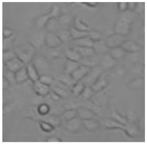
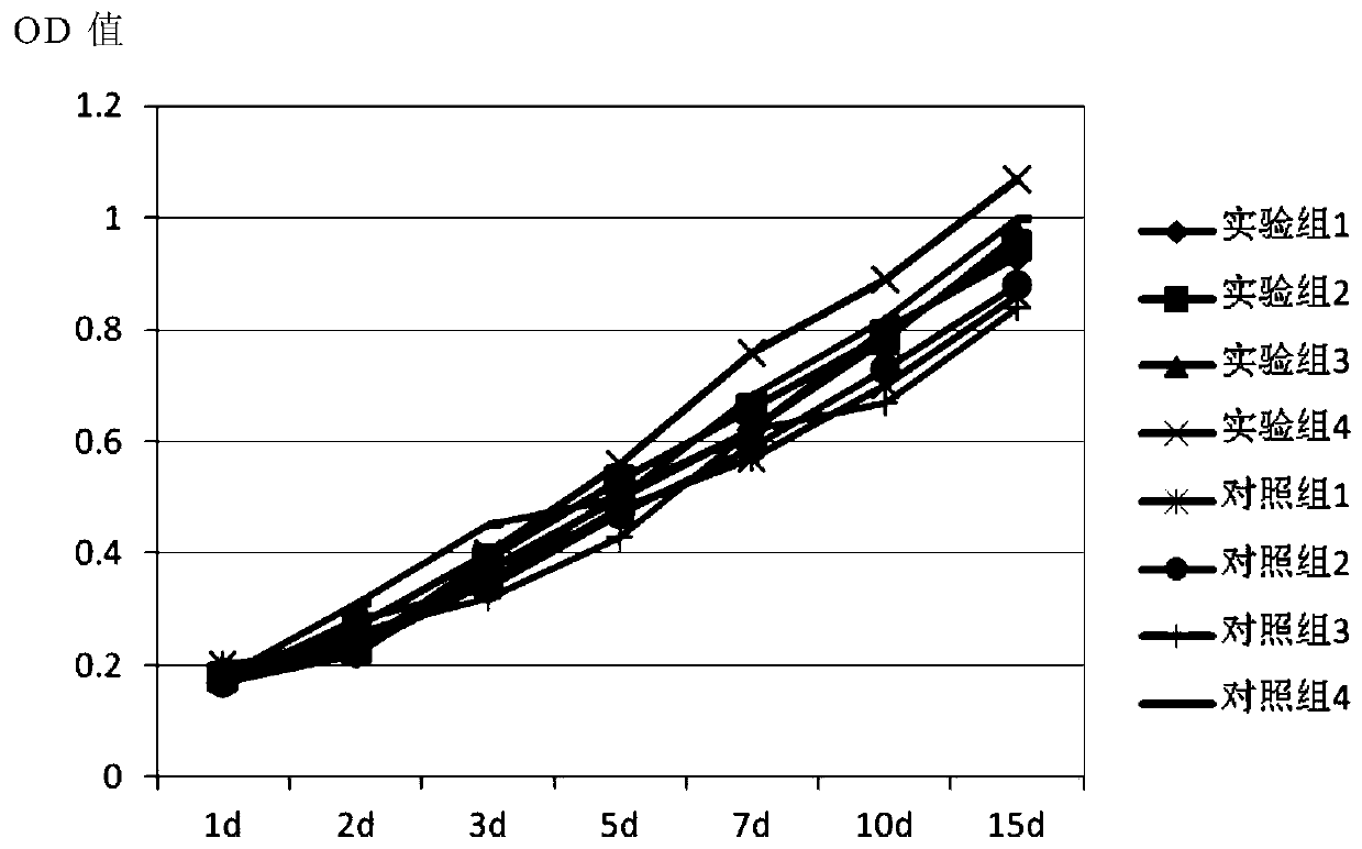
A technology of alveolar epithelium and culture method, applied in artificial cell constructs, animal cells, vertebrate cells, etc., can solve the problems of immaturity of alveolar epithelial cells in vitro, and achieve the improvement of cell viability and division ability, activity and proliferation. speed, the effect of improving the cultivation effect

Inactive Publication Date: 2019-05-17
北京昱龙摩尔国际生物医学研究院
View PDF4 Cites 3 Cited by
  • Summary
  • Abstract
  • Description
  • Claims
  • Application Information

AI Technical Summary

Problems solved by technology

However, the in vitro culture of alveolar epithelial cells is still immature

Method used


Image

  • Human alveolar epithelial cell separation and culture method
  • Human alveolar epithelial cell separation and culture method
  • Human alveolar epithelial cell separation and culture method

Examples


Embodiment 1

[0057] A method for isolating and culturing human alveolar epithelial cells, comprising the steps of:

[0058] S1: Collect lung lobe samples under sterile conditions, cut the trachea into slices, and obtain tissue fragments of alveolar epithelial tissue;

[0059] S2: An activated substrate configured for cell adherent growth, coated on a culture dish and solidified;

[0060] S3: setting an inoculation position on the solidified activated substrate, and inoculating the tissue fragments on the inoculation position, so that the tissue fragments are attached to the bottom wall of the culture dish;

[0061] S4: Add activation culture medium to the inoculated tissue fragments, place in mixed oxygen for activation culture;

[0062] S5: Transfer the activated cultured tissue fragments to a new culture dish coated with activated substrate, add the primary culture medium, and 2 Carry out primary culture in an incubator, and after 8-10 days of culture, transfer the tissue fragments to ...

Embodiment 2

[0065] A method for isolating and culturing human alveolar epithelial cells, comprising the steps provided in Example 1, wherein the specific method of step S1 is as follows:

[0066] Collect lung lobe samples under aseptic conditions, immerse in L-15 culture solution, incise the trachea, and cut into 1cm2 square slices to obtain tissue fragments of alveolar epithelial tissue.

[0067] The specific method of step S2 is as follows:

[0068] Configure the activated substrate for cell adherent growth, take 1ml and spread it on the bottom of a 6cm culture dish, and place it in CO at 36.5°C 2 Place in the incubator for 2 hours to solidify the activated substrate, and add 5 ml of activated culture solution.

[0069] The specific method of step S3 is as follows:

[0070] Use a scalpel to carve 1 cm on the edge of the bottom surface of the Petri dish 2 square, and remove the activated base to form the inoculation site; move the epithelial side of the tissue fragments into the inocu...

Embodiment 3

[0085] A method for isolating and culturing human alveolar epithelial cells, comprising the steps provided in Example 2, wherein the activated base used is the LHC-9 nutrient solution added with the following components:

[0086] Human fibronectin 0.2%; collagen 1%; fetal bovine serum 12%.


PUM


Abstract

The invention provides a human alveolar epithelial cell separation and culture method. Collected tissue is surrounded by an activated substrate to effectively grow adhering to the wall, serum ingredients can inhibit division and differentiation of alveolar epithelial cells, and therefore, the cell viability is greatly improved; tissue fragments are subjected to shake culture in a specific gaseousenvironment, damage, caused by ischemia, of the collected tissue sample can be effectively avoided, and subsequent culture operation is facilitated. Ureidohydantoin, creatine and asiaticoside added into a passage culture medium can effectively improve the cell viability and the division capacity, and therefore, cell growth and proliferation are promoted. 3T3 cells serve as the trophoderm, a good growth substrate can be provided for the primary alveolar epithelial cells, growth and division of the alveolar epithelial cells can be effectively promoted, and therefore, the culture effect is greatly improved. Through the method, the viability and the proliferation speed of the alveolar epithelial cells can be remarkably increased, and therefore, high-quality experimental materials are providedfor related research projects.

Description

technical field [0001] The invention belongs to the technical field of tissue engineering, in particular to a method for separating and culturing human alveolar epithelial cells. Background technique [0002] Airway epithelial cells are functionally active cells, which not only constitute the physical barrier of the respiratory tract, but also have the transformation and metabolism of chemical poisons or drugs, have autocrine or paracrine functions, and participate in the regulation of adjacent cell functions. Freshly isolated primary cells retain their original biological genetic characteristics and can reflect the general characteristics of cell growth in vivo, so they are suitable for research on drugs and therapies. The establishment of in vitro cell models of normal alveolar epithelial cells and diseased alveolar epithelial cells is conducive to a better understanding of the pathological changes of alveolar epithelial cells, and it is also simpler and more reliable than...

Claims


Application Information

Patent Timeline
IPC IPC(8): C12N5/071
Inventor 张晓南吴芳春吴刘兵陈虎张斌侍晓云
Owner 北京昱龙摩尔国际生物医学研究院
Learn More