Ultrasonic image and CT image fusion real-time navigation system and method

A technology of CT image and ultrasound image, applied in the field of real-time navigation system of ultrasound image and CT image fusion, can solve the problems of insufficient resolution of ultrasound image, unable to accurately describe the location and boundary of lesion area, etc., to increase ease of use, reduce Operation time, wide range of effects

Pending Publication Date: 2019-07-19
SYMBOW MEDICAL TECH
View PDF13 Cites 9 Cited by
  • Summary
  • Abstract
  • Description
  • Claims
  • Application Information

AI Technical Summary

Problems solved by technology

[0003] At present, CT image-guided percutaneous minimally invasive surgery is widely used, but due to the problem of ionizing radiation, intraoperative CT images have the disadvantage of being non-real-time. CT image-guided percutaneous minimally invasive surgery generally adopts a step-by-step puncture mode. Based on the off-line images, the clinical doctor uses the laser positioning line and the metal wire on the body surface to confirm the needle insertion path, performs the needle insertion on the same layer of the tomographic image, and scans the patient multiple times during the operation to confirm the needle tip position and path direction of the surgical needle , and the positional relationship between the surgical needle and the surrounding human tissue until the surgical instrument (surgical needle) reaches the target point
Ultrasound image-guided abdominal percutaneous minimally invasive surgery is another widely used technique. It is characterized by real-time ultrasound images and convenient equipment. However, the resolution of ultrasound images is insufficient to accurately describe the location and boundaries of the lesion area. Only suitable for percutaneous minimally invasive diagnosis and treatment operations on the superficial abdomen

Method used


Image

  • Ultrasonic image and CT image fusion real-time navigation system and method
  • Ultrasonic image and CT image fusion real-time navigation system and method

Examples


Embodiment Construction

[0030] The present invention will be described in detail below in conjunction with the accompanying drawings and embodiments.

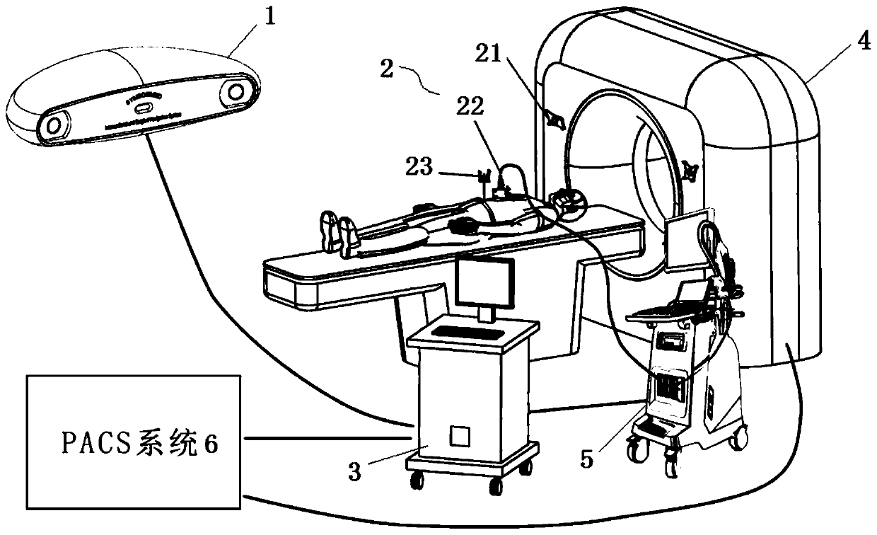
[0031] Such as figure 1 As shown, a real-time navigation system for ultrasonic image and CT image fusion provided by the present invention is applied to CT image-guided percutaneous minimally invasive operating room, and the system includes: navigation positioning sensor 1, navigation tracer 2, image processing server 3. Existing CT equipment 4, surgical instruments, ultrasound equipment 5 and PACS system 6. Wherein, the navigation positioning sensor 1 tracks the position of the ultrasonic probes of the CT equipment 4, surgical instruments, and ultrasound equipment 5 through the navigation tracer 2, and sends them to the image processing server 3; 4. The ultrasound equipment 5 is connected to the PACS system 6, and the preoperative volume images (including CT images, magnetic resonance images, PET images and CBCT (cone beam CT) images), intraoperativ...


PUM


Abstract

The invention relates to an ultrasonic image and CT image fusion real-time navigation system and method. The method is characterized by comprising the following steps: connecting all components, carrying out coordinate registration to obtain a registration matrix, and acquiring preoperative CT images or other preoperative images; performing CT image scanning to obtain CT images; according to the obtained CT images or an off-line fused image obtained by the fusion of the CT images and other preoperative images, confirming the position of the lesion tissue, confirming the needle insertion planewith the aid of an ultrasonic probe, and setting a target point / a target line; fusing the ultrasonic images into the CT images or the off-line fused image to generate a real-time fused image, and dynamically displaying the position of a surgical instrument on the real-time fused image; and updating the CT images in stages, updating the original CT images or the off-line fused image by adopting theupdated CT images, further updating the real-time fused image, and guiding percutaneous minimally-invasive surgery operations. The system can be widely applied to the field of percutaneous minimally-invasive diagnosis and treatment.

Description

technical field [0001] The invention relates to the technical field of ultrasound imaging, in particular to a real-time navigation system and method for fusion of ultrasound images and CT images. Background technique [0002] Multimodal imaging has been widely used in the field of percutaneous minimally invasive diagnosis and treatment, and the widely used images include CT, magnetic resonance, ultrasound, PET and DSA images. Various images have their own characteristics: CT images have high resolution; magnetic resonance images have advantages in soft tissue imaging; ultrasound images have the advantage of real-time; PET images are a kind of functional imaging that can reflect the metabolic information of human tissues ; DSA scanning is open, can be used for vascular imaging, and can be used as an intraoperative imaging device with flexible use. [0003] At present, CT image-guided percutaneous minimally invasive surgery is widely used, but due to the problem of ionizing r...

Claims


Application Information

Patent Timeline
Patent Type & Authority Applications(China)
IPC IPC(8): A61B34/20A61B17/34
CPCA61B34/20A61B17/3403A61B2034/2065A61B2034/2063A61B2034/2051A61B2017/3413
Inventor 肖越勇赵磊张肖费翔唐瑭张啸波边庆伟
Owner SYMBOW MEDICAL TECH
Why Eureka
Try Eureka