Method for blocking vertical transmission of avian leukosis virus and application of method
A technology of vertical transmission of avian leukosis virus, applied in the field of veterinary medicine, can solve problems such as time-consuming, high cost, and technical complexity, and achieve the effects of improving production efficiency, improving utilization rate, and reducing production links
- Summary
- Abstract
- Description
- Claims
- Application Information
AI Technical Summary
Problems solved by technology
Method used
Image
Examples
Embodiment 1
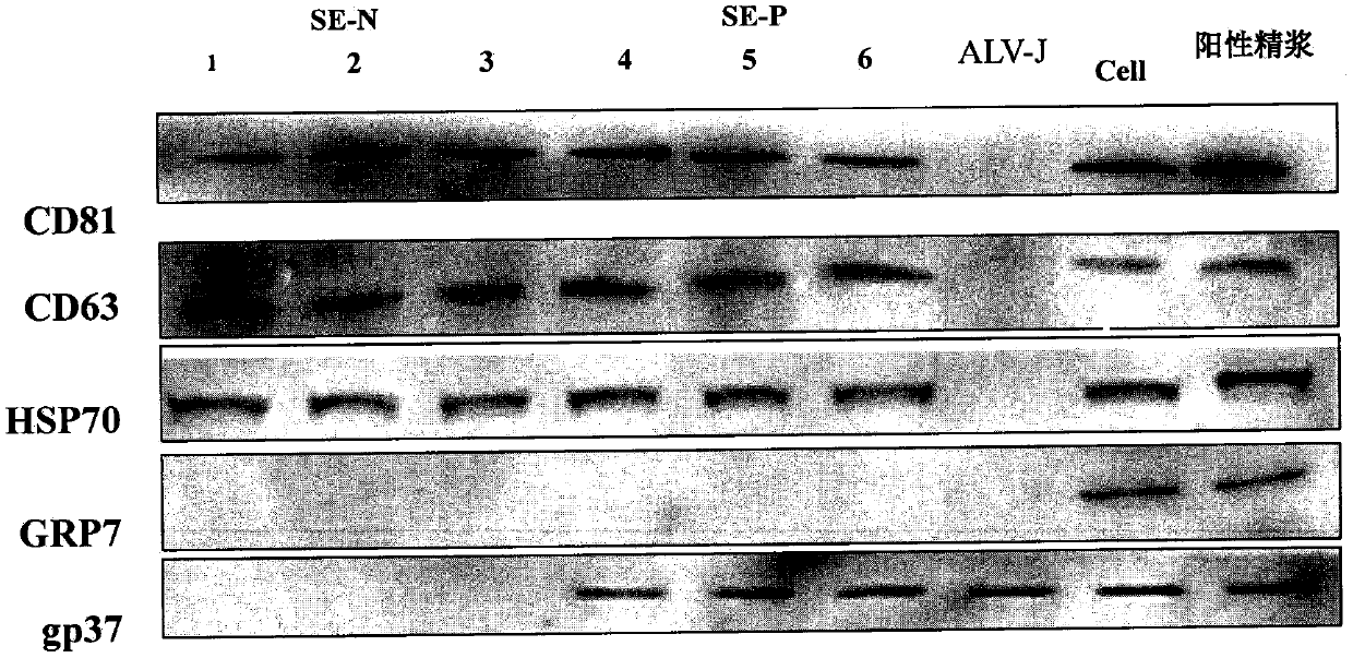
[0047] Confirmation of inclusions (ALV-J virus components) in semen exosomes
[0048] 1) Extraction and purification of exosomes in semen samples: cocks were infected with ALV-J, and blood was collected to detect viremia and p27 antibodies to confirm that the roosters were continuously infected. The semen of 3 ALV-J infected roosters and 3 healthy roosters were collected and stored at -80°C for future use. In the following examples, SEP is healthy exosomes, and SEN is positive exosomes.
[0049] Semen samples were subjected to gradient centrifugation at 4°C, respectively at 1000g, 10min; 2400g, 10min; 10000g, 30min to remove cells and cell debris, and filter macromolecular impurities with a 0.22μm filter membrane, and use the Exo-quick kit (exosome extraction Kit) mix the supernatant of the semen sample at a ratio of 4:1, incubate overnight (>12h), collect the precipitate after centrifugation at 1500g, 30min, 4°C, and resuspend with 1 / 10 the volume of the original solution in...
Embodiment 2
[0087] Infectivity of ALV-J positive chicken semen exosomes
[0088] 1) Positive exosomes infected DF-1 cells in vitro:
[0089] (1) According to 1×10 6 Inoculate DF-1 cells into a six-well plate at a ratio of / well, and the cell density reaches about 60-70%, add 50 μg and 100 μg of the purified positive semen exosomes obtained in Example 1, and incubate for 2 hours;
[0090] (2) Change 2% maintenance solution for 5 days;
[0091] (3) Cell RNA was extracted with TRIZOL Reagent for PCR detection.
[0092] Table 5: Primer sequences and parameters for ALV-J specific detection
[0093]
[0094] Table 6: Amplification reaction system
[0095]
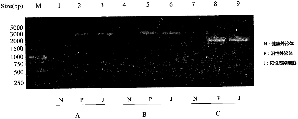
[0096] Reaction conditions: 50°C, 30min; 94°C, 5min; 94°C, 30s; 56°C, 30s; 72°C, 30s, 34 cycles; 72°C, 5min; stop the reaction at 4°C; use 1% agarose gel electrophoresis Identification.
[0097] The results show that see Image 6 , After DF-1 cells were co-incubated with positive exosomes, ALV-J virus RNA was detected in the cel...
Embodiment 3
[0101] A blocking agent for vertical transmission of ALV-J virus
[0102] (1) According to 1×10 6 Inoculate DF-1 cells to a six-well plate at a ratio of 50 μg per well, and the cell density reaches about 60-70%, and divide them into 9 groups, which are respectively the experimental group (4 Group) and the control group (group 4) that only added positive semen exosomes without adding the corresponding marker protein blocker, incubated the cells, and collected ALV-J virus RNA and protein in the cells at 12h, 24h, 36h and 72h for detection. Among them, in this example, the common marker proteins CD9, CD63, CD81 and HSP70 of exosomes were selected for blocking. The blank group was cultured pure DF-1 cells without adding positive exosomes or blockers.
[0103] see closing effect Figure 8 , compared with the control group, the addition of CD9, CD63, CD81 and HSP70 protein blockers in the experimental group can effectively reduce the proliferation of ALV-J virus in cells, and red...
PUM
 Abstract
Description
Claims
Application Information
 - R&D
- Intellectual Property
- Life Sciences
- Materials
- Tech Scout
- Unparalleled Data Quality
- Higher Quality Content
- 60% Fewer Hallucinations
Browse by: Latest US Patents, China's latest patents, Technical Efficacy Thesaurus, Application Domain, Technology Topic, Popular Technical Reports.
© 2025 PatSnap. All rights reserved.Legal|Privacy policy|Modern Slavery Act Transparency Statement|Sitemap|About US| Contact US: help@patsnap.com


