
Chromatographic analysis column, kit and device for detecting prosthetic joint infection

A chromatographic analysis and kit technology, applied in measurement devices, analytical materials, material separation, etc., can solve the problems of inability to achieve immediate diagnosis, no effective solution, insufficient sensitivity, etc., and achieve improved stability and separation. Good, the effect of improving sensitivity

Active Publication Date: 2022-01-07
北京谷海天目生物医学科技有限公司
View PDF7 Cites 0 Cited by
  • Summary
  • Abstract
  • Description
  • Claims
  • Application Information

AI Technical Summary

Problems solved by technology

These indicators still have some deficiencies in the specificity and sensitivity of diagnosing PJI: 1) Bacterial culture is one of the "gold" standards for diagnosing PJI, but the positive rate of bacterial culture is about 60% to 70%, and the sensitivity is not high enough
2) Counting and classification of white blood cells in synovial fluid is an effective method for diagnosing PJI, but after joint replacement in patients with systemic inflammatory arthritis, this index will also increase, and the specificity is not strong
3) Existing laboratory diagnostic methods cannot achieve immediate diagnosis (Point of Care Testing, POCT, or bedside diagnosis)
[0004] Therefore, how to detect PJI more accurately, there is still no effective solution in the prior art

Method used


Image

  • Chromatographic analysis column, kit and device for detecting prosthetic joint infection
  • Chromatographic analysis column, kit and device for detecting prosthetic joint infection
  • Chromatographic analysis column, kit and device for detecting prosthetic joint infection

Examples


Embodiment 1

[0097] The heavy isotope-labeled standard peptides shown in SEQ ID NO:1, SEQ ID NO:2, SEQ ID NO:3 and SEQ ID NO:4 were prepared with a final concentration of 100fmol of peptides, then shake and mix, centrifuge for 3 minutes on a 12000g centrifuge, and take the supernatant for later use;

[0098] Extract protein from 40 joint fluid samples from individuals with prosthetic joints, and then perform enzymatic hydrolysis. Take 100 μg of joint fluid proteolysis peptides from each sample and add 100 μL of loading buffer, shake and mix to make a joint The liquid peptide solution is ready for use;

[0099] Take 1 μL of joint fluid peptide solution, and add 1 μL of 100 fmol heavy isotope-labeled standard peptide solution to it, then add 3uL of loading buffer, mix well, and place on a 96-well loading plate for chromatographic injection.

[0100] Mix the synthesized heavy-labeled peptides in an equimolar ratio, and mix the individual peptides at the following concentrations: 250fmol, 62....

Embodiment 2

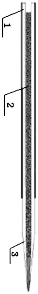
[0123] The difference between Example 2 and Example 1 is that in the chromatographic analysis column, the length of the chromatographic column tube is 150 mm, the outer diameter is 360 μm, and the inner diameter is 150 μm, and the filler is C18 reverse phase filler with a particle size of 1.9 μm. Meanwhile, the chromatographic separation time was 75 minutes.

Embodiment 3

[0125] The difference between Example 3 and Example 1 is that in the chromatographic analysis column, the length of the chromatographic column tube is 300 mm, the outer diameter is 360 μm, and the inner diameter is 150 μm, and the filler is C18 reverse phase filler with a particle size of 1.9 μm. Meanwhile, the chromatographic separation time was 150 minutes.

[0126] Both Examples 2 and 3 can effectively identify the target peptide, and carry out targeted quantitative analysis of the target peptide, and the results obtained can also well distinguish whether the prosthetic joint is infected or not (due to the data in Example 1 There is no significant difference and will not be specifically listed here). However, considering the timeliness of clinical sample detection and the cost of mass spectrometry detection, the detection conditions in Example 1 in this application are the best application conditions.

[0127] From the above description, it can be seen that the above-menti...


PUM

PropertyMeasurementUnit
lengthaaaaaaaaaa
lengthaaaaaaaaaa
lengthaaaaaaaaaa

Abstract

The invention provides a chromatographic analysis column, a kit and a device for detecting prosthetic joint infection. The chromatographic analysis column comprises: a chromatographic column tube, a filler filled in the chromatographic column tube; and a chromatographic column tip integrated with the chromatographic column tube, and the inner diameter of the chromatographic column tube is 100-200 μm. The use of a relatively wide inner diameter can enable the chromatographic column tube to maintain the column pressure within the normal pressure range when increasing the chromatographic elution flow rate, which improves the stability of chromatographic separation; it is also conducive to increasing the sample loading and improving Sensitivity of mass spectrometry detection. In addition, the integrated chromatographic analysis column tip has no tailing phenomenon during elution, and the separation of proteins is better. Therefore, when the chromatographic analysis column improved in this application is used to detect PIJ, the separation degree of the peptide segment is better and the stability is higher, thereby making the quantitative result of the target protein more accurate.

Description

technical field [0001] The invention relates to the field of prosthetic joint infection detection, in particular to a chromatographic analysis column, a kit and a device for detecting prosthetic joint infection. Background technique [0002] Prosthetic joint infections (PJI) is recognized as one of the most serious complications after joint replacement. Although the incidence of PJI is not high, it has not been significantly reduced since the application of antibiotics. The reported incidence of hip and shoulder PJI is approximately 1%, knee PJI incidence is approximately 2%, and elbow PJI incidence is approximately 9%. For arthroplasty, the incidence of PJI is even as high as 40%; in addition, 33% of the complications leading to the failure of total ankle arthroplasty are PJI. [0003] According to the diagnostic guidelines proposed by the American Academy of Orthopedic Surgeons, the Infectious Diseases Society, and the Musculoskeletal Infection Society, most PJIs can be c...

Claims


Application Information

Patent Timeline
Patent Type & Authority Patents(China)
IPC IPC(8): G01N30/88G01N30/38
CPCG01N30/88G01N30/38G01N2030/8831
Inventor 秦钧陈继营汪宜丁琛宋雷李睿李恺刘明伟
Owner 北京谷海天目生物医学科技有限公司
Learn More