Biosensor and method for detecting microcystin
A technology of biosensors and microcystins, which is applied to instruments, measuring devices, scientific instruments, etc., can solve the problems of difficulty in realizing in-situ real-time detection of microcystins, high testing costs, complicated pretreatment, etc., and achieves a simple structure. , reduce the difference between batches, the effect of good application prospects
- Summary
- Abstract
- Description
- Claims
- Application Information
AI Technical Summary
Problems solved by technology
Method used
Image

Examples
Embodiment 1
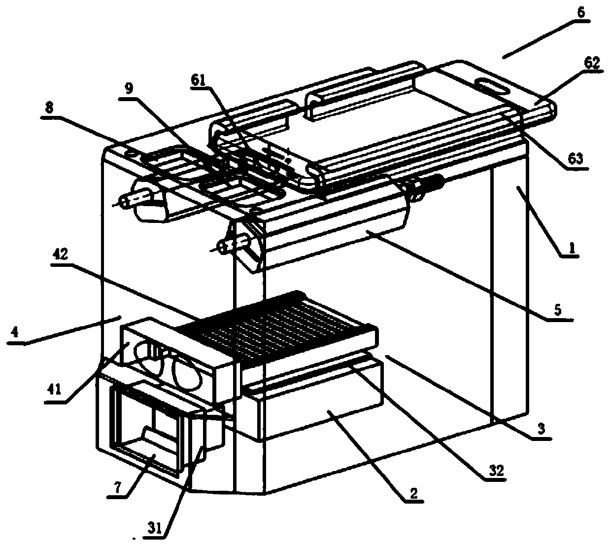
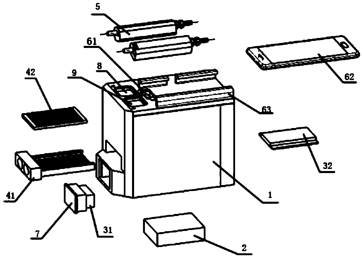
[0037] A biosensor for the detection of microcystins, such as figure 1 , figure 2 As shown, it includes a biosensor housing 1, a power supply module 2 located inside the biosensor housing 1, an incubation module 3, an immune reaction module 4, a fluorescence excitation module 5 and a signal acquisition module 6 located at the top of the biosensor housing 1 ; The power supply module 2 is arranged at the bottom of the biosensor housing 1, and is connected to the incubation module 3 and the fluorescence excitation module 5 respectively; Connection composition; one side of the biosensor housing 1 is provided with a temperature display screen 7, and the temperature display screen 7 is connected to the intelligent temperature controller 31; Composed of chips 42; the fluorescent excitation module 5 is located on the inside of the top of the biosensor housing 1, and is composed of an LED array, an excitation filter, and an emission filter. The LED array and the top of the biosensor ...
Embodiment 2
[0041] A method for detecting microcystins, comprising the steps of:
[0042] S1. Preparation of microcystin immunosensor based on nano-magnetic beads;
[0043] S2. Using the above-mentioned biosensor to measure the fluorescence intensity produced by the biosensor under the standard series of microcystin concentrations, and draw a linear relationship curve between the microcystin concentration and the fluorescence intensity;
[0044] S3. Use the above-mentioned biosensor to detect the microcystin in the water, and calculate the concentration of the microcystin in the sample by using the linear relationship curve drawn in step S2.
Embodiment 3
[0046] Example 3 Preparation of Microcystin Immunosensor Based on Nano Magnetic Beads
[0047] 1. Activation of magnetic beads
[0048] Take 500 μL of magnetic beads and place them in a 1.5mL EP tube, place them in a magnetic separation rack, enrich the magnetic beads, and remove the supernatant; add 1 mL of 1mM hydrochloric acid solution pre-cooled at 4°C, vortex for 15 seconds, and place in a magnetic separation rack for enrichment Magnetic beads, remove supernatant.
[0050] Add 500 μL of protein solution (MC-LR antigen at a concentration of 250 μg / mL), vortex for 30 s, and mix well. Put the EP tube on the vortex oscillator, adjust to the lowest gear, automatic mode, and oscillate for 1-1.5h.
[0051] Wherein, the ratio of the nano magnetic beads to the microcystin antigen coated on the surface is 1:1.
[0052] 3. Separation
[0053] Use a magnetic separation rack to enrich the magnetic beads and save the flow-through.
[0054] 4. Cleaning...
PUM
 Abstract
Description
Claims
Application Information
 - R&D
- Intellectual Property
- Life Sciences
- Materials
- Tech Scout
- Unparalleled Data Quality
- Higher Quality Content
- 60% Fewer Hallucinations
Browse by: Latest US Patents, China's latest patents, Technical Efficacy Thesaurus, Application Domain, Technology Topic, Popular Technical Reports.
© 2025 PatSnap. All rights reserved.Legal|Privacy policy|Modern Slavery Act Transparency Statement|Sitemap|About US| Contact US: help@patsnap.com