
Method and system for stable and quantitative analysis of ultrasound contrast images

A contrast-enhanced ultrasound and image stabilization technology, applied in image analysis, ultrasound/sonic/infrasound image/data processing, ultrasound/sonic/infrasonic Permian technology, etc. Unreasonable and stable feature quantization parameters, etc., to achieve the effect of improving operation speed, stability and high accuracy

Pending Publication Date: 2019-09-03
SICHUAN PROVINCIAL PEOPLES HOSPITAL
View PDF7 Cites 4 Cited by
  • Summary
  • Abstract
  • Description
  • Claims
  • Application Information

AI Technical Summary

Problems solved by technology

The determination of the region of interest generally relies on the doctor's naked eye observation and experience judgment, which has a large personal subjectivity
However, in the existing quantification technology of contrast imaging, due to external uncontrollable factors (such as the continuous movement of the probe, the breathing of the human body, etc.), the tissue movement is deformed and the region of interest disappears, which in turn causes the parameter quantification index to jitter violently and the peak value is extremely low. , so that the visual effect of the fitted evaluation curve is not intuitive, and the quantitative parameters of the obtained angiographic features are also unreasonable and stable, and cannot provide stable and reliable basic data information for the quantitative analysis and disease diagnosis of medical workers.

Method used


Image

  • Method and system for stable and quantitative analysis of ultrasound contrast images
  • Method and system for stable and quantitative analysis of ultrasound contrast images
  • Method and system for stable and quantitative analysis of ultrasound contrast images

Examples


Embodiment Construction

[0020] In the following, the present invention will be further described in detail in conjunction with the accompanying drawings and embodiments, so as to make the purpose, technical solutions and advantages of the present invention more clear. It should be understood that the specific embodiments described here are only used to explain the present invention, not to limit the present invention.

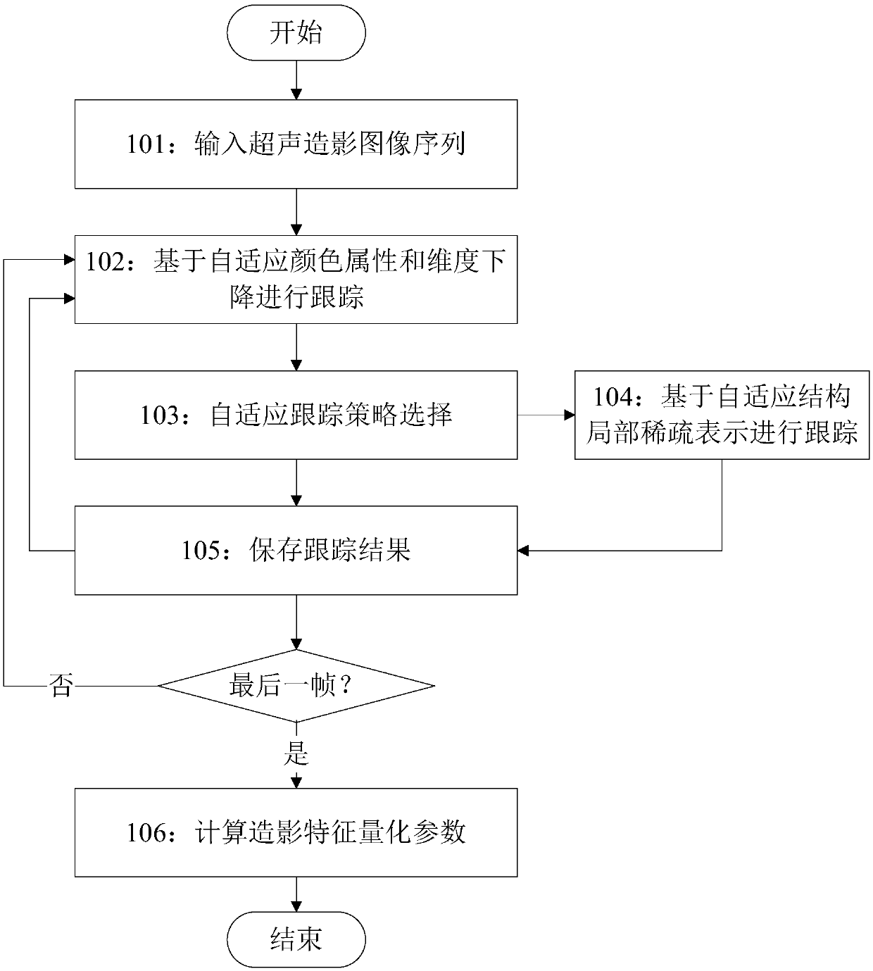
[0021] Such as figure 1 As shown, the method for stable quantitative analysis of contrast-enhanced ultrasound images according to an embodiment of the present invention includes the following steps:

[0022] Step 101: Acquiring a sequence of ultrasound-enhanced images with multiple frames, as well as position and size information of the target area;

[0023] Specifically, a contrast-enhanced ultrasound image sequence including multiple frames may be acquired by reading from a memory or directly inputting through a contrast-enhanced ultrasound imaging probe, and displaying one frame (...


PUM


Abstract

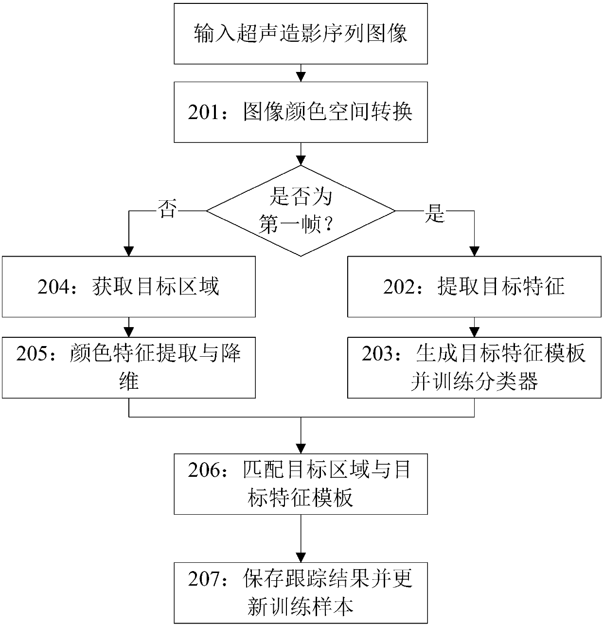
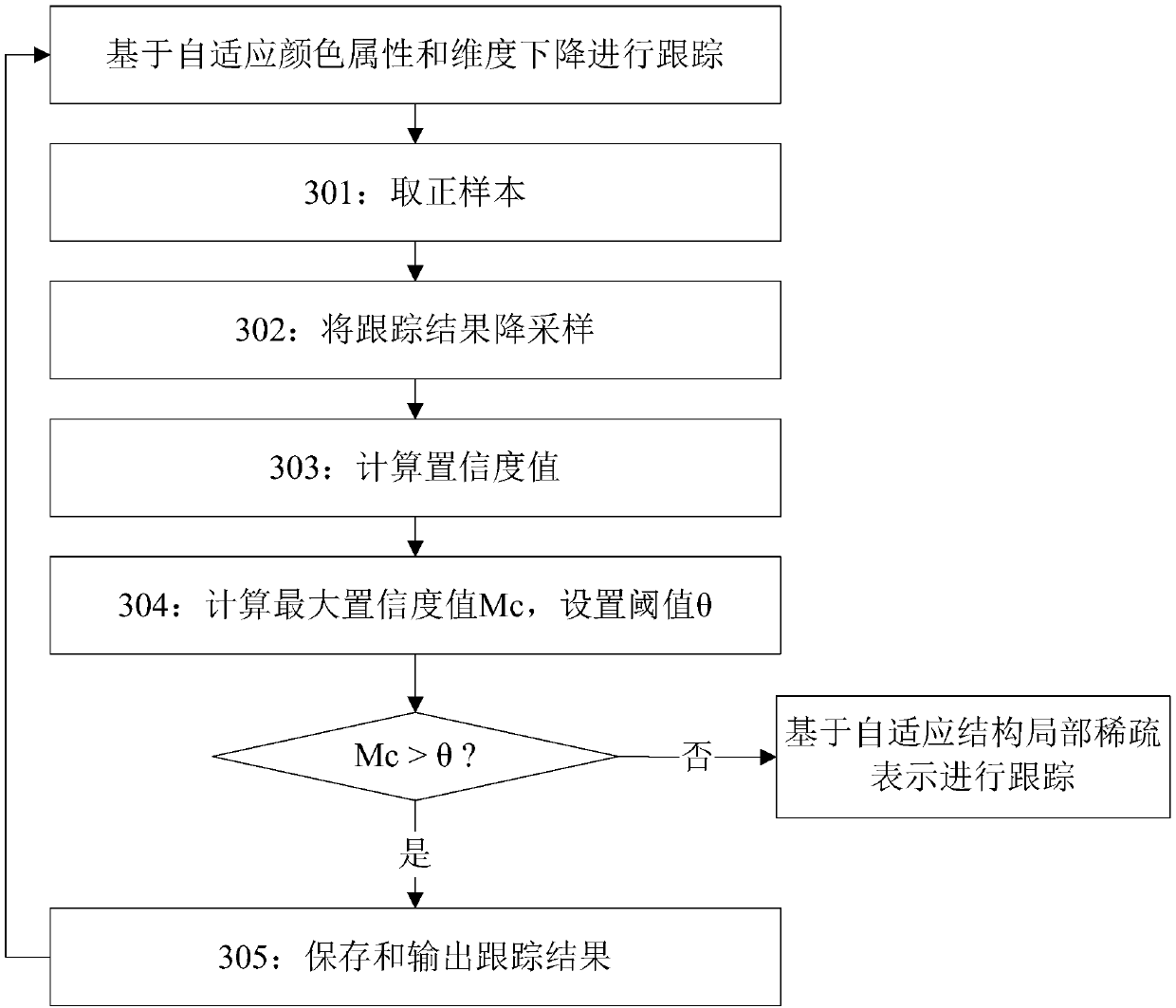
The invention discloses a method and a system for stable and quantitative analysis of ultrasound contrast images, which can solve the problems of target deformation, scale change and irregular motionin the tracking of points of interest of the ultrasound contrast image, and the stability and the accuracy of obtained contrast characteristic quantization parameters are high. The method comprises the steps of tracking a current frame in an ultrasound contrast image sequence based on adaptive color attributes and dimension reduction, and obtaining a tracking result comprising position and size information of a target area in a next frame; performing adaptive tracking strategy selection based on confidence evaluation, and performing whether a tracking result is stored and a next frame continues to be tracked based on adaptive color attributes and dimensionality decline or tracking again based on adaptive structure local sparse representation so as to obtain a tracking result is determined;and after all frames are processed, calculating a radiography intensity value according to the stored tracking result data, forming an original time intensity curve TIC, performing curve fitting, andcalculating radiography characteristic quantification parameters.

Description

technical field [0001] The invention relates to the technical field of contrast-enhanced ultrasound imaging analysis, in particular to a method and system for stable quantitative analysis of contrast-enhanced ultrasound images. Background technique [0002] Contrast-enhanced ultrasound imaging technology uses contrast agents to enhance backscattered echoes and diagnose lesions by observing blood perfusion. Contrast-enhanced ultrasonography can provide basic information for the diagnosis of heart, liver, kidney, pancreas, peripheral blood vessels, thyroid, breast, prostate, uterus and other solid organs, and can also be further used to assist in the evaluation of kidney and liver transplantation effects. [0003] In contrast-enhanced ultrasonography diagnosis, doctors generally obtain the diagnosis results based on the contrast agent perfusion of the lesion and the doctor's personal experience. However, conclusions drawn by doctors based on experience are sometimes subjectiv...

Claims


Application Information

Patent Timeline
IPC IPC(8): G06T7/00A61B8/08
CPCG06T7/0012A61B8/5215G06T2207/10016G06T2207/10132
Inventor 罗俊龚勋易心一许新
Owner SICHUAN PROVINCIAL PEOPLES HOSPITAL
Learn More