

Image contrast enhancement method, system and device
An image comparison and image technology, applied in the field of medical image processing and application, can solve problems such as increasing the complexity of clinical applications, reduce work and training burden, improve clinical efficiency, and enhance morphological features.
- Summary
- Abstract
- Description
- Claims
- Application Information
AI Technical Summary
Problems solved by technology
Method used
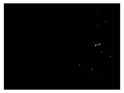
Image


Examples
Embodiment Construction
[0044] The present invention will be described in detail below in conjunction with the implementations shown in the drawings, but it should be noted that these implementations are not limitations of the present invention, and those of ordinary skill in the art based on the functions, methods, or structural changes made by these implementations Equivalent transformations or substitutions all fall within the protection scope of the present invention.
[0045] In the present invention, the optical fiber microendoscope system uses an optical fiber bundle as a probe for microscopic imaging. Because the light transmittance of a single optical fiber core and the cladding in the optical fiber bundle is different, the image collected will have obvious pixelation (brightness and darkness change) ), reducing the system resolution. The commonly used depixelization method is Gaussian filtering. Although this method is simple, fast and effective, it will blur the entire image and affect the...
PUM
 Abstract
Description
Claims
Application Information