Construction method, storage medium and computing equipment of sinoatrial node virtual physiological tissue
A construction method and organizational technology, applied in the field of translational medicine, can solve problems such as lack of experimental data, lack of systematic understanding of sinus node function, and obstacles to the understanding of sinoatrial node function, so as to overcome moral pressure, overcome animal experiment defects, reduce The effect of time and money spent
- Summary
- Abstract
- Description
- Claims
- Application Information
AI Technical Summary
Problems solved by technology
Method used
Image
Examples
Embodiment 1
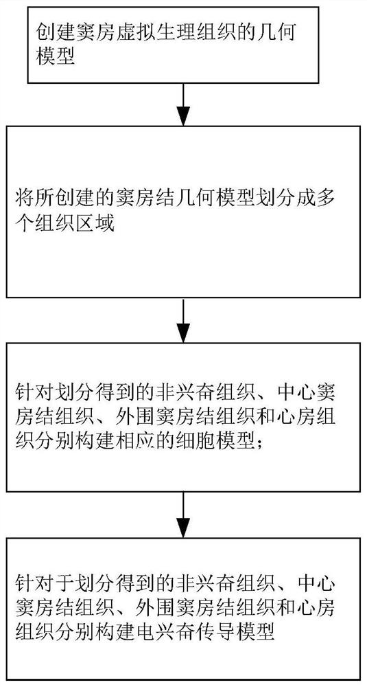
[0072] The invention discloses a method for constructing sinoatrial node virtual physiological tissue, which is implemented in a computer, such as figure 1 shown, including the following steps:
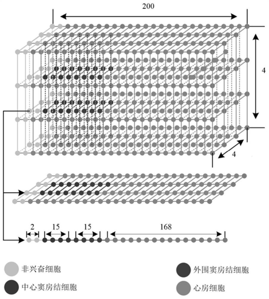
[0073] S1. Create a geometric model of virtual physiological tissue; in this embodiment, as Figure 2a and 2b As shown, the geometric model of the virtual physiological tissue is a cuboid, and the number of nodes in length, width and height are 200, 4 and 4 respectively; there is a certain distance between every two adjacent nodes in length, width and height , called the spatial step; where each node represents a cell.
[0074] S2. Dividing the created sinoatrial node virtual physiological tissue geometric model into multiple regions, including a non-exciting tissue region, a central sinoatrial node tissue region, a peripheral sinoatrial node tissue region, and an atrial tissue region;
[0075] Such as Figure 2a and 2b As shown, in this embodiment, in the sinoatrial node virtual...
Embodiment 2
[0127] This embodiment discloses a storage medium, which stores a program. When the program is executed by a processor, the method for constructing the sinoatrial node virtual physiological tissue described in Embodiment 1 is realized, specifically as follows:
[0128] Create a geometric model of the virtual physiological tissue of the sinoatrial node;
[0129] Divide the created sinoatrial node geometric model into multiple regions, including non-exciting tissue region, central sinoatrial node tissue region, peripheral sinoatrial node tissue region and atrial tissue region;
[0130] Corresponding cell models were constructed for the divided non-excitatory tissue, central sinus node tissue, peripheral sinus node tissue and atrial tissue:
[0131]
[0132] Among them, for non-exciting tissues, the first cell model was constructed using a sinoatrial node cell model that does not contain L-type calcium ion current: Among them, C m1 Indicates non-exciting tissue membrane cap...
Embodiment 3
[0152] The invention discloses a computing device, including a processor and a memory for storing executable programs of the processor. When the processor executes the program stored in the memory, the method for constructing the sinoatrial node virtual physiological tissue in Embodiment 1 is realized. Specifically as follows:
[0153] Create a geometric model of the virtual physiological tissue of the sinoatrial node;
[0154] Divide the created sinoatrial node geometric model into multiple regions, including non-exciting tissue region, central sinoatrial node tissue region, peripheral sinoatrial node tissue region and atrial tissue region;
[0155] Corresponding cell models were constructed for the divided non-excitatory tissue, central sinus node tissue, peripheral sinus node tissue and atrial tissue:
[0156]
[0157] Among them, for non-exciting tissues, the first cell model was constructed using a sinoatrial node cell model that does not contain L-type calcium ion cu...
PUM
 Abstract
Description
Claims
Application Information
 - R&D
- Intellectual Property
- Life Sciences
- Materials
- Tech Scout
- Unparalleled Data Quality
- Higher Quality Content
- 60% Fewer Hallucinations
Browse by: Latest US Patents, China's latest patents, Technical Efficacy Thesaurus, Application Domain, Technology Topic, Popular Technical Reports.
© 2025 PatSnap. All rights reserved.Legal|Privacy policy|Modern Slavery Act Transparency Statement|Sitemap|About US| Contact US: help@patsnap.com


