Cardiac medical image processing method, processing device, processing system and medium

A medical image and processing method technology, applied in image data processing, ultrasonic/sonic/infrasonic image/data processing, image enhancement, etc., can solve the problem of delineating the edge of the myocardial region, difficulty in tracking, and the inability of the instrument to automatically identify the myocardial region, etc. , to achieve the effect of reducing the workload

Active Publication Date: 2021-06-11
SHANGHAI YIZHI HEALTHCARE TECH CO LTD
View PDF5 Cites 0 Cited by
  • Summary
  • Abstract
  • Description
  • Claims
  • Application Information

AI Technical Summary

Problems solved by technology

Due to the semi-automatic operation, the instrument cannot automatically identify the myocardial area, and the doctor needs to manually outline it in the first frame of the image. At the same time, the instrument’s built-in outline curve tracking function cannot accurately outline the edge of the myocardial area in the subsequent dynamic video, so it cannot be accurately detected. Change track of myocardial speckle
It is necessary to select several pairs of myocardial spots and detect their average trajectory to calculate the myocardial strain rate. However, for patients with non-uniform myocardial thickness such as hypertrophic cardiomyopathy, only the distance change between limited pairs of myocardial spots cannot truly reflect the overall myocardial strain rate. deformation
Furthermore, the shape of myocardial speckles in each frame of image is not exactly the same, and there are often some changes, and even some myocardial speckles may not appear in some frames, which brings difficulties to tracking

Method used


Image

  • Cardiac medical image processing method, processing device, processing system and medium
  • Cardiac medical image processing method, processing device, processing system and medium
  • Cardiac medical image processing method, processing device, processing system and medium

Examples


Embodiment Construction

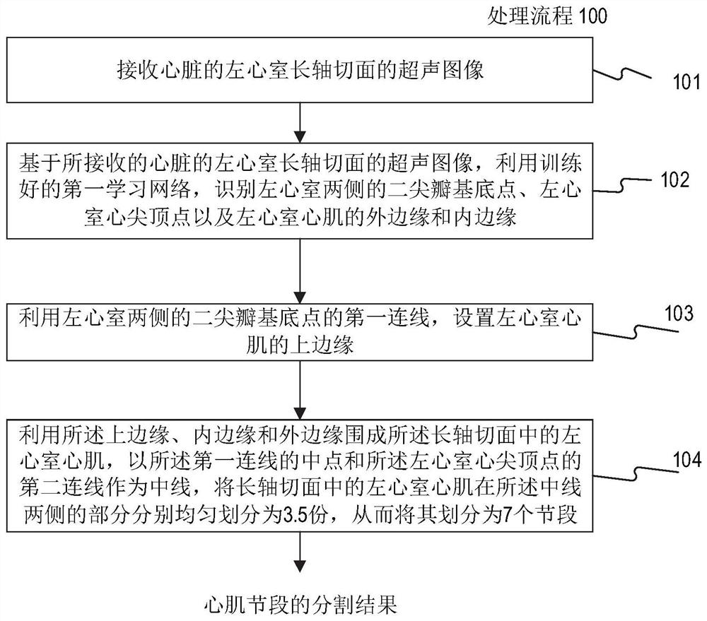
[0029] figure 1A flowchart showing a method 100 for processing cardiac medical images according to the first embodiment of the present disclosure. As used herein, medical images of the heart may include images of the heart region acquired with various imaging modalities including, but not limited to, ultrasound imaging, functional MRI (e.g., fMRI, DCE-MRI, and diffusion MRI) , cone beam CT (CBCT), helical CT, positron emission tomography (PET), single photon emission computed tomography (SPECT), X-ray imaging, optical tomography, fluorescence imaging, and radiotherapy portal imaging, etc. The processing method of the present disclosure will be described below by taking an ultrasonic image of the heart as an example, but it should be known that the processing method can be flexibly applied to medical images of the heart in various other imaging modalities besides the ultrasonic image of the heart.

[0030] Such as figure 1 As shown, the processing method 100 begins at step 10...


PUM


Abstract

The present disclosure relates to a processing method, a processing device, a processing system and a medium for cardiac medical images. The method includes: receiving a cardiac medical image of a long-axis view of the left ventricle; using a trained first learning network to identify the mitral valve base points on both sides of the left ventricle, the apex apex, and the outer edge and inner edge of the left ventricular myocardium; The first connecting line of the mitral valve base point, set the upper edge; use the upper edge, inner edge and outer edge to enclose the left ventricular myocardium, and use the midpoint of the first connecting line and the second connecting line of the apex of the left ventricle as the midline , the parts of the left ventricular myocardium on both sides of the midline are evenly divided into n parts, thereby dividing it into 2n segments. The processing method, processing device, processing system and medium of the cardiac medical image can automatically, accurately and quickly delineate and identify various myocardial regions, greatly reducing the workload of doctors, and its automatic identification can continue to benefit from the labeling of myocardial regions after training training data set.

Description

technical field [0001] The present disclosure generally relates to image processing and analysis. More specifically, the present disclosure relates to a processing method, a processing device, a processing system and a medium for cardiac medical images. Background technique [0002] The traditional parameter for evaluating overall myocardial function is mainly left ventricular ejection fraction. This single-dimensional parameter cannot comprehensively evaluate the overall myocardial function, let alone accurately reflect the diastolic and contractile movements of the local myocardium. Damage is often a gradual process from local to overall. [0003] Myocardial strain (MS) refers to the deformation of the myocardium during the cardiac cycle, which refers to the percentage change of the size of the myocardium in a certain dimension relative to the initial myocardial length or the state of no stress, and can be used to interpret the elongation of the myocardium Or shorten, th...

Claims


Application Information

Patent Timeline
Patent Type & Authority Patents(China)
IPC IPC(8): G06T7/00G06T7/13G06T7/66G06T7/68A61B8/08
CPCA61B8/5223G06T7/0012G06T2207/10132G06T2207/20081G06T2207/20084G06T2207/30048G06T7/13G06T7/66G06T7/68
Inventor 石磊倪浩郑永升印宏坤杨俊沈庆
Owner SHANGHAI YIZHI HEALTHCARE TECH CO LTD
Why Eureka
Try Eureka