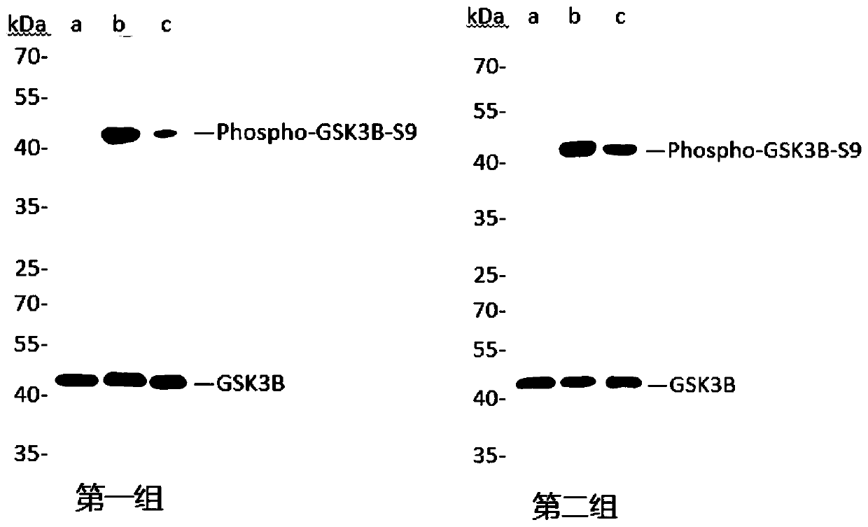
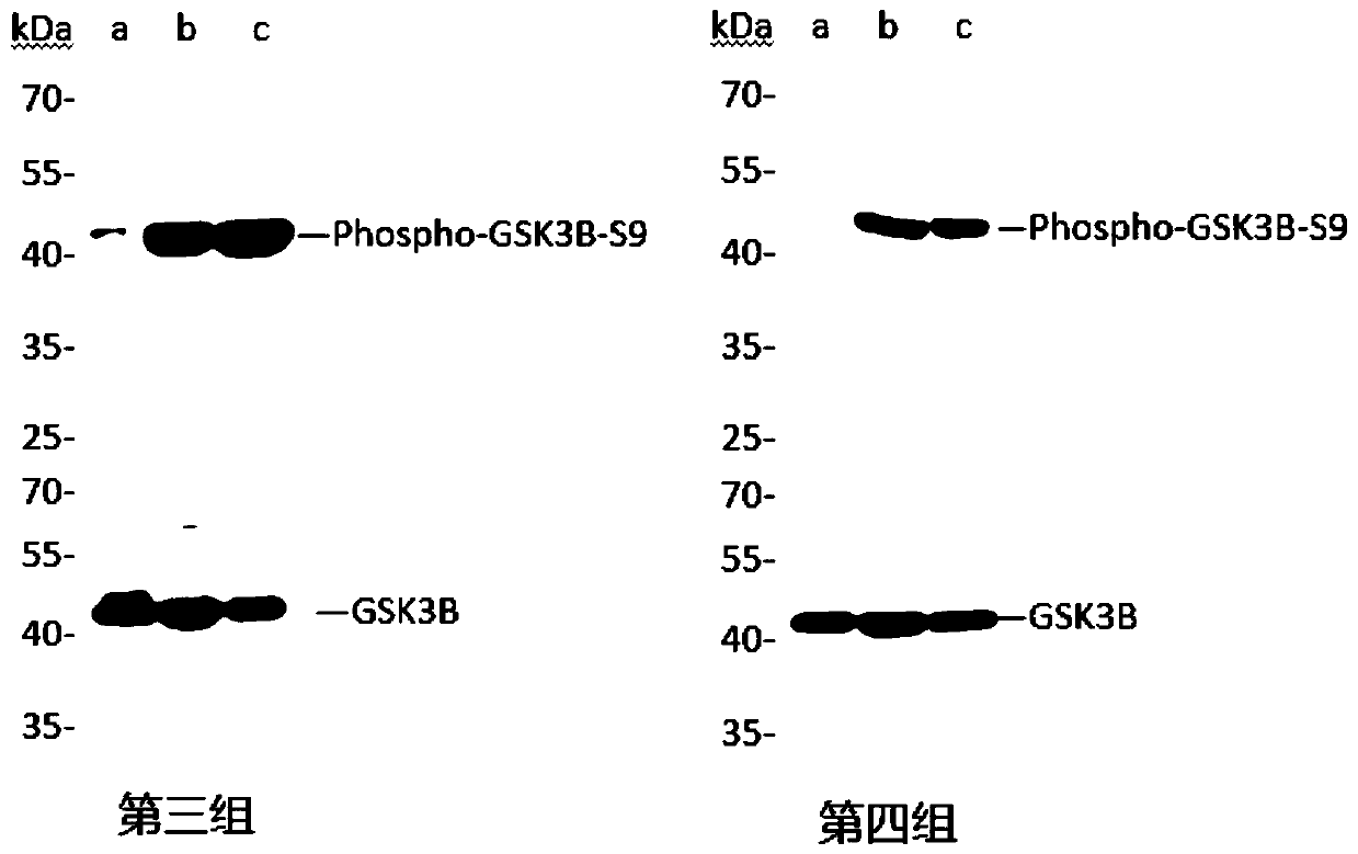
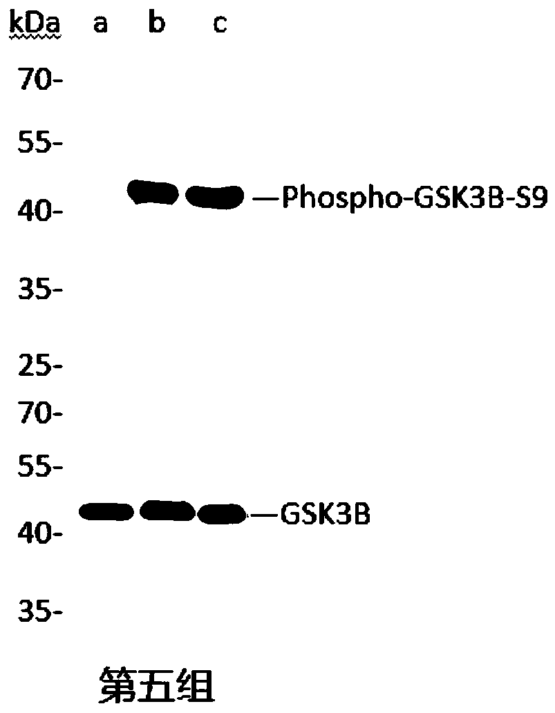
Method for preparing ATP in vitro phosphorylation sample

A phosphorylation and phosphatase inhibitor technology, applied in the field of ATP phosphorylation sample preparation in vitro, can solve the problems of time-consuming, cumbersome experiments, and inability to obtain phosphorylated proteins

Inactive Publication Date: 2020-01-17
WUHAN AIBO TAIKE BIOTECH CO LTD
View PDF6 Cites 2 Cited by
  • Summary
  • Abstract
  • Description
  • Claims
  • Application Information

AI Technical Summary

Problems solved by technology

The disadvantage is that a broad spectrum of phosphorylated proteins cannot be obtained, and a single drug can only phosphorylate a small part of the protein....

Method used


Image

  • Method for preparing ATP in vitro phosphorylation sample
  • Method for preparing ATP in vitro phosphorylation sample
  • Method for preparing ATP in vitro phosphorylation sample

Examples


Embodiment 1

[0037] 1. Cell culture:

[0038] 1.1. Cell recovery

[0039] Take out the frozen HeLa cells and immediately put them into a 42°C constant temperature water bath. Shake slightly to thaw the cells quickly. After the cells are completely thawed, take out the cryovial and place the cryovial into a 50mL centrifuge tube. Balance, 1000rpm / min. Centrifuge for 5 min. After centrifugation, take out the cryovial, spray some alcohol into the ultra-clean workbench, light the alcohol lamp, open the T25 cell culture flask, add 4 mL of DMEM medium (Boehringer Ingelheim / BI); unscrew the cryovial and discard the supernatant Add 1 mL of culture medium and pipette the cells into a suspension, transfer them to a T25 culture flask, mark the cell type, date, and the name of the cultivator on the culture flask, shake it gently and place it in an incubator for culture .

[0040] 1.2. Cell Passaging

[0041] 1.2.1. Adherent cells

[0042] 1) Observe under a microscope. When the cell coverage i...


PUM


Abstract

A Method for preparing an ATP in vitro phosphorylation sample comprises the following steps of culturing cells; collecting the cells to obtain cellular precipitation; extracting cell protein: placingthe cellular precipitation collected in the step S2 in ice and adding a cell lysis buffer for cell ultrasonic pyrolysis, and performing low-temperature centrifugation after ice incubation to obtain aprotein extract, wherein the cell ultrasonic pyrolysis lasts 10min; adding ATP to the protein extract described in the step S3, making the final concentration of ATP as 5mM, performing constant temperature water bath for 1h at 30 DEG C after uniform mixing to obtain the protein extract; performing protein denaturation after protein concentration measurement; and diluting the sample for storage at-80 DEG C. The invention does not need to stimulate cells with numerous drugs to obtain one phosphorylated protein after another. In this system, we can achieve the phosphorylation of almost all proteins, which plays a very good role in the verification of phosphorylated antibodies.

Description

technical field [0001] The present invention designs a sample preparation method, more specifically, relates to a sample preparation method for ATP phosphorylation in vitro. Background technique [0002] The research and development of phosphorylation in vivo is relatively mature. There are two main types of phosphorylation of proteins in vivo: [0003] One is to phosphorylate one, several or a certain type of proteins on the pathway according to the signal transduction pathway in the organism, so this method is often accompanied by the study of the signal pathway, and it is necessary to find suitable substances to stimulate cells. , so that the cell body is activated or closed on this signaling pathway, so as to obtain the desired phosphorylated protein. [0004] The second is based on the activity of protein kinases required for protein phosphorylation. In this way, it is also necessary to find suitable substances to activate or inactivate related protein kinases to obtai...

Claims


Application Information

Patent Timeline
IPC IPC(8): G01N1/28C12P21/00
CPCC12P21/00G01N1/28
Inventor 黄长青李军陈文
Owner WUHAN AIBO TAIKE BIOTECH CO LTD
Why Eureka
Try Eureka