
Esophageal cancer pathological image labeling method

A technology for pathological images and esophageal cancer, applied in medical images, image enhancement, image analysis, etc., can solve the problems of long training period for pathologists, energy consumption, and waste of medical resources, so as to avoid manual feature selection process and save time and cost , the simple effect of the model

Pending Publication Date: 2020-02-21
SHANDONG COMP SCI CENTNAT SUPERCOMP CENT IN JINAN
View PDF10 Cites 5 Cited by
  • Summary
  • Abstract
  • Description
  • Claims
  • Application Information

AI Technical Summary

Problems solved by technology

The interpretation of medical images requires the accumulation of professional experience for a long time, and the training cycle of pathologists is relatively long, and esophageal cancer lesions occur in the basal layer of esophageal epithelial tissue, close to the junction of epithelial tissue and mesenchymal tissue, so pathologists are required to strictly Defining the contours of epithelial tissue with fine lines is labor-intensive and a huge waste of medical resources

Method used


Image

  • Esophageal cancer pathological image labeling method
  • Esophageal cancer pathological image labeling method
  • Esophageal cancer pathological image labeling method

Examples


Embodiment Construction

[0048] The present invention will be further described below in conjunction with the accompanying drawings and embodiments.

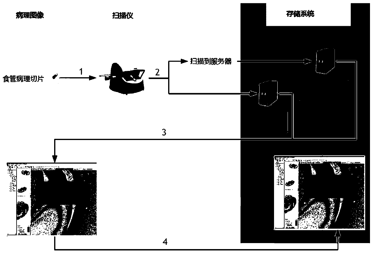
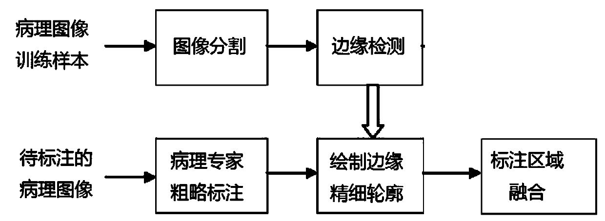
[0049] Such as figure 1 As shown, the flow chart of the method for annotating esophageal cancer pathological images of the present invention is given, figure 2 A flow chart of building an epithelial tissue contour detection model in the present invention is given, which is realized through the following steps:

[0050] a). Image staining correction, staining correction processing is performed on the H&E stained esophagus pathological image, and the color difference between the pathological images due to the uneven staining produced during the section staining process is reduced;

[0051] In this step, the method for image dyeing and correction is as follows: first, according to Lambert-Beer's law, the color value is converted into an optical density value, and using singular value decomposition, the hematoxylin Haematoxylin and eosin Eosin used for pa...


PUM


Abstract

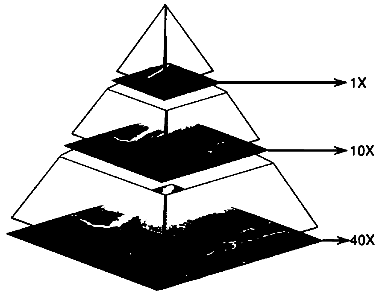
The esophageal cancer pathological image labeling method comprises the following steps: a) performing dyeing correction processing on an esophageal pathological image subjected to H & E dyeing; b) labeling expert canceration areas; c) mapping the canceration area outline marked by the expert into a pathological image of 40X; d) constructing an epithelial tissue contour detection model; d-1) marking whether pixel points belong to an epithelial region, an interstitial tissue or an irrelevant blank region; (d-2) constructing an end-to-end convolutional neural network model; and e) fusing the labeling areas. According to the method, the epithelial tissue contour is automatically drawn in the labeling process according to the characteristic that the esophageal cancer morbidity area occurs in the epithelial tissue basal layer area, so that the time cost of expert labeling is greatly saved. The method only aims at esophageal pathological section image modeling; only the edge of the epithelialtissue is detected, the model is relatively simple, operation is rapid, epithelial boundary detection has obvious advantages in detection precision, meanwhile, the method automatically learns effective features and expressions, the complex manual feature selection process is avoided, and the actual application requirements can be met.

Description

technical field [0001] The present invention relates to a method for annotating pathological images of esophageal cancer, and more specifically, to a method for annotating pathological images of esophageal cancer, which can convert rough contours marked by experts into precise contours. Background technique [0002] Esophageal cancer is a common malignant tumor in life, which seriously affects human health. At present, it is more and more difficult to rely on experts to screen and diagnose esophageal cancer pathological slides as the number of patients increases. The combination of medical imaging and artificial intelligence is a relatively new branch and industrial hotspot in the field of digital medicine. The application of artificial intelligence in the medical field has become a trend. The application of big data-driven artificial intelligence to build a computer-aided diagnosis system for esophageal cancer and relieve the work pressure and intensity of doctors is the d...

Claims


Application Information

Patent Timeline
Patent Type & Authority Applications(China)
IPC IPC(8): G06K9/32G06K9/46G06N3/04G06T7/13G16H30/20
CPCG06T7/13G16H30/20G06T2207/30004G06V10/25G06V10/44G06V10/56G06N3/045Y02A90/10
Inventor 牛春阳
Owner SHANDONG COMP SCI CENTNAT SUPERCOMP CENT IN JINAN
Learn More