Target extraction method of symmetric supervision model based on mixed loss function

A technology of mixed loss and loss function, applied in image data processing, 3D image processing, instruments, etc., can solve the problems of manpower and material resources, low efficiency, etc., and achieve the effect of improving efficiency and solving manpower and material resources.

Active Publication Date: 2020-03-10
HARBIN INST OF TECH
View PDF7 Cites 3 Cited by
  • Summary
  • Abstract
  • Description
  • Claims
  • Application Information

AI Technical Summary

Problems solved by technology

[0003] The purpose of the present invention is to solve the problems that the existing left atrium extraction method consumes a lot of manpower and material res

Method used


Image

  • Target extraction method of symmetric supervision model based on mixed loss function
  • Target extraction method of symmetric supervision model based on mixed loss function
  • Target extraction method of symmetric supervision model based on mixed loss function

Examples


specific Embodiment approach 1

[0029] Specific implementation mode 1: The specific process of the target extraction method based on the symmetric supervision model of the mixed loss function in this implementation mode is as follows:

[0030] Step 1. Obtain cardiac magnetic resonance image data and label them;

[0031] Step 2. Preprocessing the labeled cardiac MRI image data obtained in step 1, the preprocessing includes operations such as obtaining two-dimensional slices, cropping, scaling, and data normalization;

[0032] Step 3, using the cardiac MRI image data preprocessed in step 2 as the input of the symmetric supervision model;

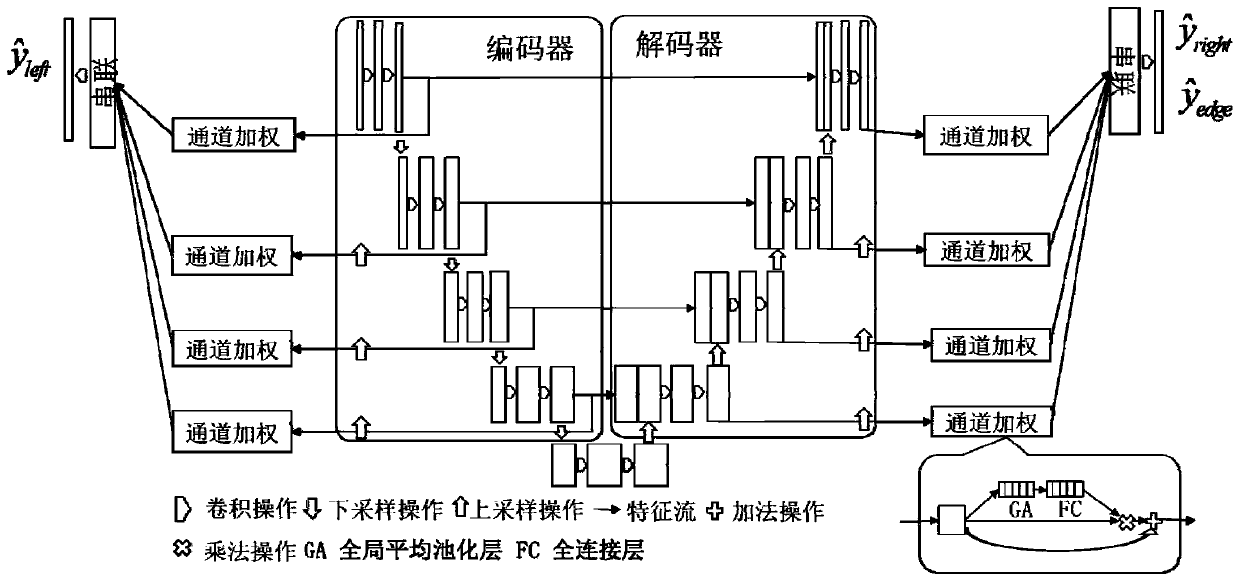
[0033] The symmetric supervised model consists of two parts: an encoder and a decoder. The encoder includes an input layer, a convolutional layer, a normalization layer, a maximum pooling layer, and an output layer. The decoder includes a convolutional layer, a deconvolutional layer, and a normalization layer. Oneization layer, Dropout layer and output layer;

[0034] The ...

specific Embodiment approach 2

[0041] Specific embodiment two: the difference between this embodiment and specific embodiment one is that in the step 1, cardiac MRI image data is acquired and marked; the specific process is:

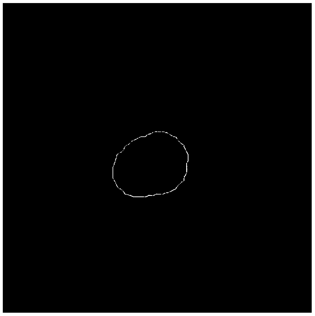
[0042] Step 11, manually mark the left atrium area and the background area, wherein the left atrium area is marked as 1, and the background area is marked as 0, and this type of mark is stored as the label of the extraction model;

[0043] Step 12, use the Canny operator to extract boundaries on the left atrium region and background region binary images extracted in step 11, mark the boundaries as 1, and mark other regions as 0, and store such marks as labels for boundary detection.

[0044] Other steps and parameters are the same as those in Embodiment 1.

specific Embodiment approach 3

[0045] Specific embodiment three: the difference between this embodiment and specific embodiment one or two is that in the step 2, the labeled cardiac MRI image data obtained in step 1 is preprocessed, and the preprocessing includes obtaining two-dimensional slices, cutting , scaling, and data normalization operations; the specific process is:

[0046] In the training phase, random rotation within the specified range and horizontal and vertical flip operations will be performed on the labeled cardiac MRI image data obtained in step 1 to achieve data amplification to avoid overfitting;

[0047] Two-dimensional slices are extracted from the marked cardiac MRI image data obtained in step 1 through the long axis (the MRI is scanned in the direction from head to toe of the person, in fact, a slice is scanned at each moment, and then reconstructed into 3D It is stored as a 3D array, and this extraction is converted into a 2D slice in the direction of the long axis according to the o...


PUM


Abstract

The invention discloses a target extraction method of a symmetric supervision model based on a mixed loss function, and relates to a target extraction method. The invention aims to solve the problemsthat the existing left atrium extraction method consumes a large amount of manpower and material resources, has human differences and is low in efficiency. The method comprises the following steps: 1,acquiring heart nuclear magnetic resonance image data, and labeling; 2, preprocessing the marked heart nuclear magnetic resonance image data obtained in the step 1; 3, obtaining a trained symmetric supervision model, and storing the trained symmetric supervision model; 4, predicting the preprocessed heart nuclear magnetic resonance image data by adopting the symmetric supervision model trained inthe step 3, outputting the probability that each pixel is judged as the left atrium, and setting a probability threshold to binarize a result; and 5, reconstructing the binarized slice result obtained in the step 4 into three-dimensional volume data according to the inverse operation of the step 2 to finish the extraction of the left atrium. The method is applied to the field of target extraction.

Description

technical field [0001] The present invention relates to a target extraction method. Background technique [0002] Medical image processing is a new discipline and technology that has developed rapidly with the development and maturity of computer technology and the advancement of clinical diagnostic technology. Nowadays, medical image processing technology is more and more widely used in clinical practice as an auxiliary tool for doctors. . The left atrium is an important part of the human heart, and its structure, size and shape are important factors to identify the physiological state of the human body. The clinical method for extracting the left atrium is still at the stage of manual extraction by doctors using software (such as CVI42, Circle cardiac imaging). This type of method has large human-subjective differences, and requires relevant personnel with professional knowledge to be responsible Work. The extraction efficiency is also relatively low, which greatly incr...

Claims


Application Information

Patent Timeline
IPC IPC(8): G06T15/00G06T7/12
CPCG06T15/00G06T7/12G06T2207/10088G06T2207/30048G06T2207/10012
Inventor 王宽全刘亚淑骆功宁王玮张恒贵
Owner HARBIN INST OF TECH
Why Eureka
Try Eureka