Detection method for biological efficacy of mesenchymal stem cells

A stem cell biology, stem cell technology, applied in the field of detection of the biological efficacy of mesenchymal stem cells, can solve the problems of many steps, inconvenient detection, instability, etc., and achieve the effects of good detection means, simple operation, and accurate quantification.

Pending Publication Date: 2020-03-31
北京贝来生物科技有限公司
View PDF3 Cites 4 Cited by
  • Summary
  • Abstract
  • Description
  • Claims
  • Application Information

AI Technical Summary

Problems solved by technology

However, in actual operation, it is found that this method has the following disadvantages: 1. Fresh blood from healthy people needs to be drawn for each test, which brings inconvenience to the test; 2. The human peripheral blood lymphocytes taken for each test come from different donors, Therefore, there is instability between batches; 3. This method can provide qualitative results of the biological efficacy of mesenchymal stem cells, but cannot give quantitative indicators; 4. This method needs to recover and culture mesenchymal stem cells before seeding to 96 Orifice plate, then co-culture with peripheral blood lymphocytes (add PHA and other stimulators) for 48-72 hours, and finally detect the level of TNF-α in the culture medium. The whole experiment takes 6-7 days, with many steps and complicated procedures

Method used


Image

  • Detection method for biological efficacy of mesenchymal stem cells
  • Detection method for biological efficacy of mesenchymal stem cells
  • Detection method for biological efficacy of mesenchymal stem cells

Examples


Embodiment 1

[0057] Example 1 Isolation and cultivation of human umbilical cord-derived mesenchymal stem cells (hUC-MSC)

[0058] Umbilical cord tissue was obtained from a cooperative hospital, and the donor had signed an informed consent. hUC-MSCs were isolated, cultured and expanded from umbilical cord tissue by tissue climbing method.

[0059] The specific method is as follows: select umbilical cord tissue from a healthy donor over 15 cm, cut the umbilical cord tissue into small sections with a diameter of 1-1.5 mm, and inoculate 56 pieces / bottle evenly into T75 culture bottles. Place the culture bottle containing the umbilical cord tissue at 37° C. in a 5% CO incubator for 5-6 hours. After the incubation, add 5 ml of complete cell culture medium (DMEM / F12 (DF12) culture solution plus 10 % fetal bovine serum (FBS)), add 10ml of complete culture medium after overnight culture, after that, change the culture medium every 3-5 days, and carefully observe the level of cells climbing out of ...

Embodiment 2

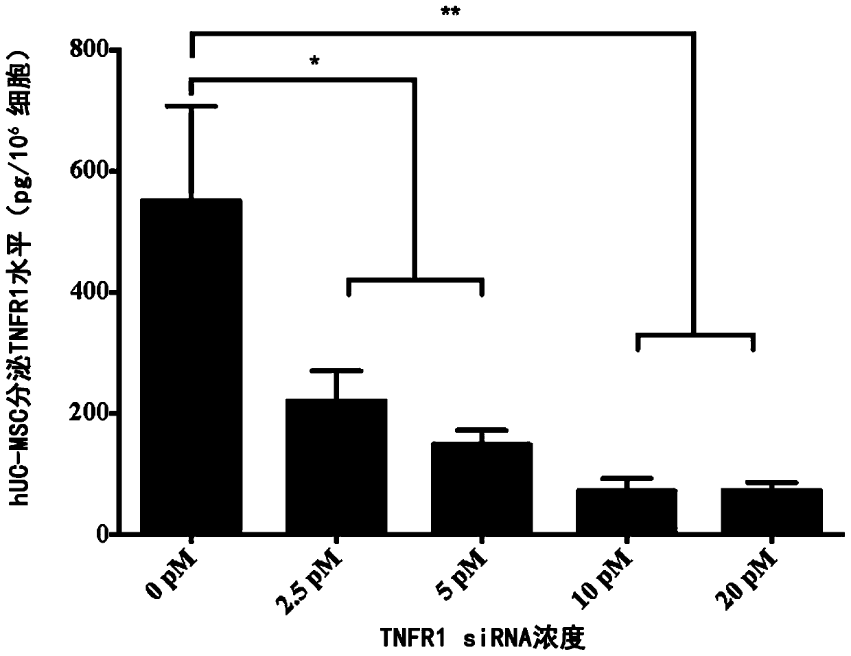
[0061] Example 2 TNFRI siRNA treatment of hUC-MSC, detection of cell secreted TNFRI level

[0062]TNFRI siRNA (s14266) was dissolved in sterile nuclease-free water to a 10 μM preservation solution. siRNA (s14266, Thermofisher company) and liposome transfection reagent ( RNAi-MAX reagent) for Medium dilution. Each concentration of siRNA was mixed with the same volume of RNAi-MAX for 5 minutes at room temperature, and the final concentration of TNFRI siRNA was adjusted to 0, 2.5, 5, 10 or 20 pM. Prepare parallel plates for cell counting.

[0063] hUC-MSC cells produced under GMP environment from 4 different donor sources were used for analysis: UC001, UC002, UC005 and UC006. The hUC-MSCs were recovered, counted, and seeded in 24-well cell culture plates at a seeding density of 3×10⁴ cells / well. Each batch of cells was cultured in one culture plate (4 culture dishes in total). After culturing the cells overnight, TNFRI siRNA (s14266) at a final concentration of 0, 2.5, 5,...

Embodiment 3

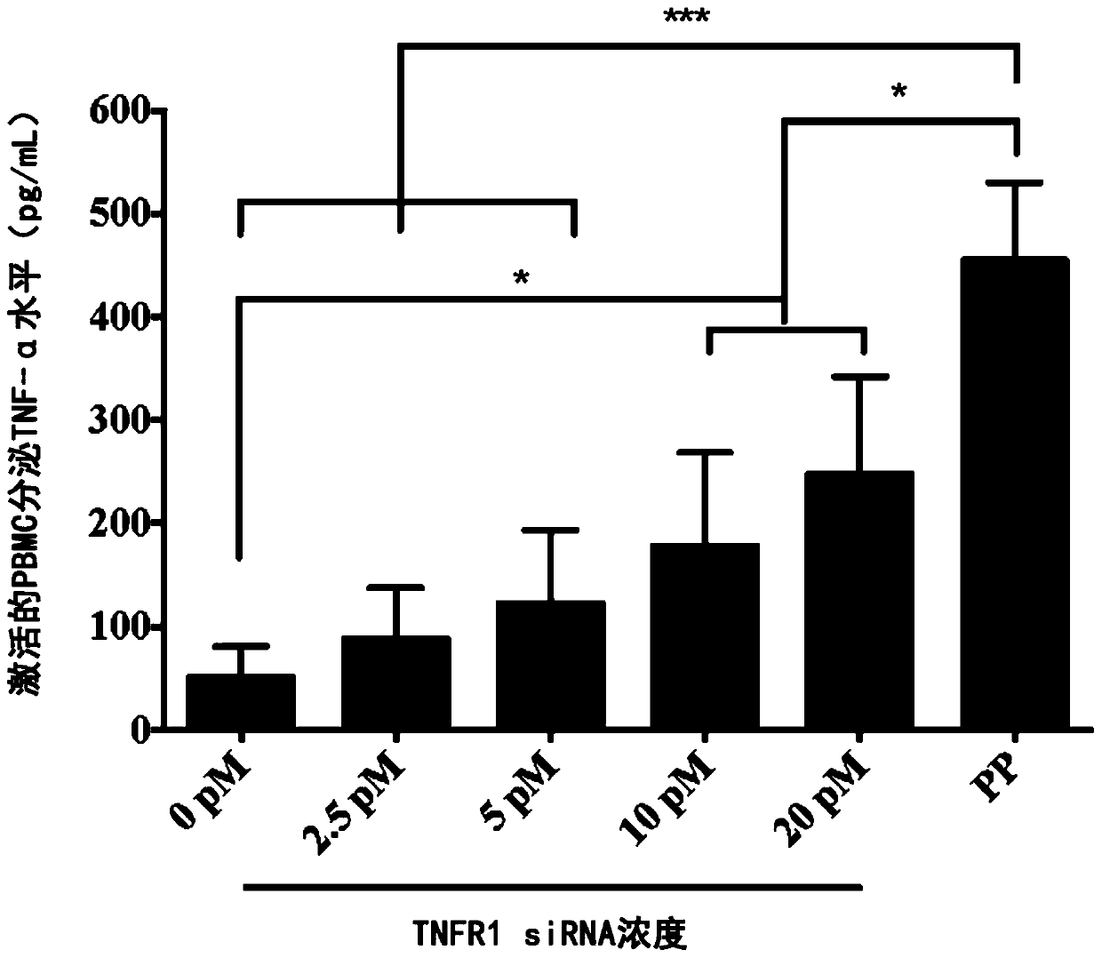
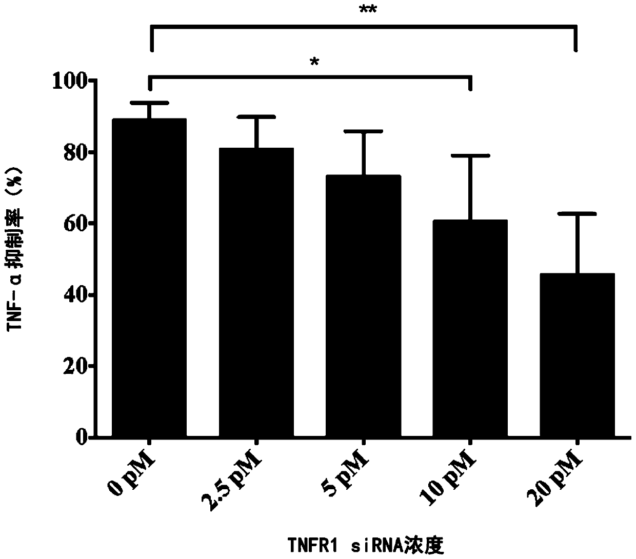
[0067] Example 3 co-culture of hUC-MSC and PBMC, detection of TNF-α

[0068] Human peripheral blood was obtained from healthy donors. Peripheral blood lymphocytes were separated from blood using Ficoll-Paque PLUS reagent, then the cells were washed twice with DPBS, and finally resuspended with RPMI-1640 complete culture medium. After counting PBMC cells, adjust the cell concentration to 1×10⁷ cells / ml. PBMC cells were preactivated with anti-CD3 antibody (final concentration: 1 μg / ml) and anti-CD28 antibody (final concentration: 1 μg / ml), and the preactivated PBMCs will be used for co-culture immediately.

[0069] Count the number of hUC-MSCs (UC001, UC002, UC005 and UC006) cultured in parallel 24-well plates, remove the culture medium from the hUC-MSCs (cells treated with different concentrations of TNFRI siRNA) in each experimental group, and add 200 μL RPMI-1640 to each well The culture medium was completed; the preactivated PBMCs were added to each hUC-MSC experimental we...


PUM

PropertyMeasurementUnit
diameteraaaaaaaaaa

Abstract

The invention provides a detection method for the biological efficacy of mesenchymal stem cells. The method is characterized by detecting the level of expression secretion type TNFRI in a culture supernatant of the mesenchymal stem cells by an ELISA process to determine the strength of the immunosuppression ability of the cells. The method is easy to operate, high in pertinence and accurate in quantification, and provides a better detection means of the biological efficacy of the mesenchymal stem cells.

Description

technical field [0001] The present invention relates to a detection technology of mesenchymal stem cells, in particular to a method for detecting the biological efficacy of mesenchymal stem cells, which is used as the mesenchymal stem cell by detecting the level of anti-inflammatory factors in the culture supernatant of mesenchymal stem cells. Quality Control Standards for Biological Potency. Background technique [0002] Tumor necrosis factor-α (TNF-α) is a major immunomodulatory and pro-inflammatory factor that is thought to play a role in the pathology of autoimmune diseases such as rheumatoid arthritis and ulcerative colitis due to its upregulation. important role in the mechanism. In rheumatoid arthritis, TNF-α is mainly produced by synovial macrophages in RA synovium and stimulates fibroblast proliferation and lymphocyte activity. The process of TNF-α generation begins with the binding of ligands and cell surface Toll-like receptors (TLRs), which stimulates signal tr...

Claims


Application Information

Patent Timeline
Patent Type & Authority Applications(China)
IPC IPC(8): C12Q1/02C12N5/071A61K35/28A61P37/02
CPCA61K35/28A61P37/02C12N5/0665G01N33/5091
Inventor 刘广洋刘拥军张晨亮李欣米一苗丽马静
Owner 北京贝来生物科技有限公司
Why Eureka
Try Eureka