A kind of modeling method and animal model and its application

An animal model and modeling technology, applied in the field of medicine, can solve problems such as injury, unstable intraocular pressure control, and anterior capsule puncture

Active Publication Date: 2021-08-20
GENERAL HOSPITAL OF PLA
View PDF6 Cites 0 Cited by
  • Summary
  • Abstract
  • Description
  • Claims
  • Application Information

AI Technical Summary

Problems solved by technology

The bigger disadvantage of this model is that it is prone to complications, such as anterior capsule perforation, cataract, corneal injury and infection, etc.
[0010] Although there are many ways to make animal models of ocular hypertension, most of the current animal models of glaucoma have high requirements for equipment and operators, unstable intraocular pressure control, and most of them are invasive, causing great interference to intraocular tissues, etc. shortcoming

Method used


Image

  • A kind of modeling method and animal model and its application
  • A kind of modeling method and animal model and its application
  • A kind of modeling method and animal model and its application

Examples


Embodiment 1

[0057] A method for making a mouse model of ocular hypertension, comprising the following steps:

[0058] (1) After general anesthesia, C57BL / 6J mice were placed in lateral position;

[0059] (2) Topical anesthesia is given to the surface of the eyeball, and the pupil is dilated;

[0060] (3) Take 3 cm of silicone rubber cerclage with a width of 3 mm used in human external scleral cerclage, and prepare to make a cerclage ring with a diameter smaller than that of the mouse eyeball.

[0061] (4) Cut off the wedge-shaped ends of the opposite ends of the cerclage to make the appearance of the end of the cerclage as a sharp point, and make a soft silicone transparent sleeve with a width of 1mm.

[0062] (5) The two ends of the cerclage are respectively inserted into a soft silicone transparent sleeve with a width of 1mm, and the diameter of the cerclage ring and the length of both ends are adjusted;

[0063] (6) The cerclage loop made is ligated on the sclera surface 1 mm behind ...

Embodiment 2

[0076] After the rabbit was fully anesthetized, the surface of the eyeball was given surface anesthesia with obcaine, and the silicone rubber cerclage used in scleral cerclage was taken to make a cerclage loop. The two ends of the cerclage were inserted into soft silicone rings respectively, and were tied outside the sclera 1.5mm behind the limbus of the rabbit. After fixing the position, the cerclage was tightened and the cerclage was ligated. The intraocular pressure was measured by a tonometer, and then fixed The ends of the cerclage in the collar are non-returnable.

Embodiment 3

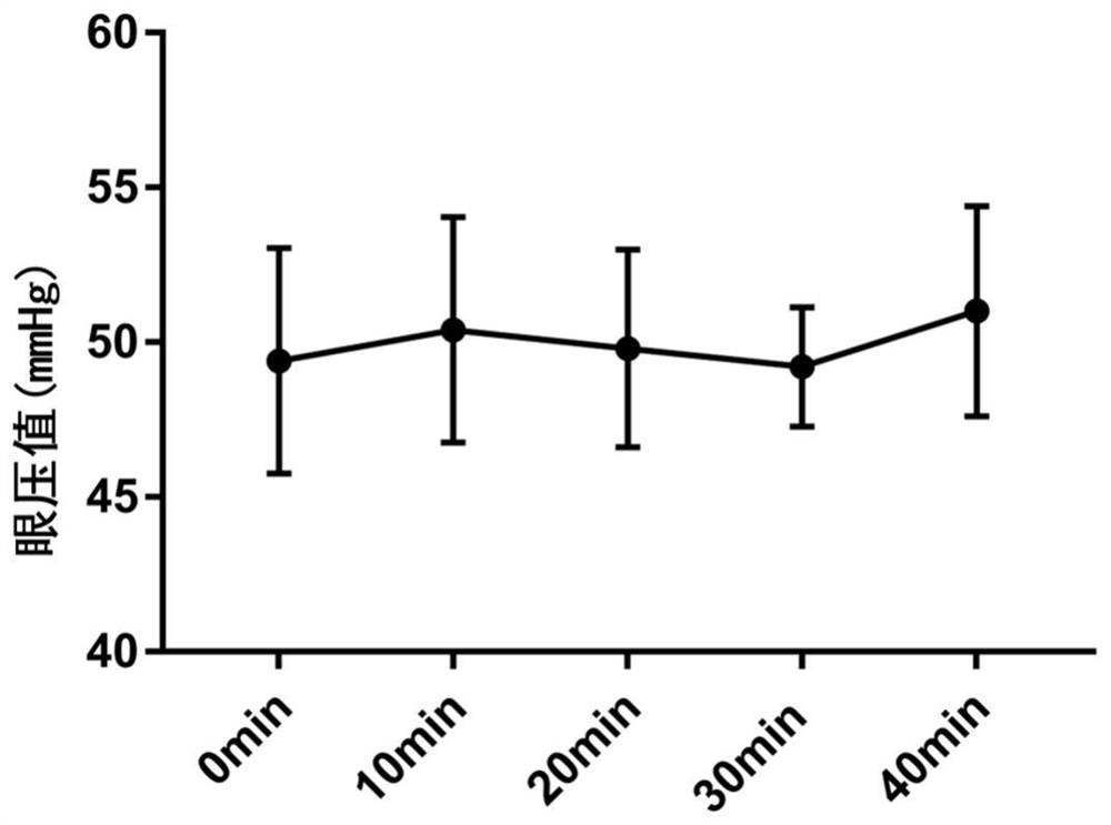
[0077] Embodiment 3: intraocular pressure measurement of model animals

[0078] Tonopen tonometer measurement: The tonometer is calibrated before each use. After the calibration is successful, the intraocular pressure measurement is completed by the same person alone. Before the measurement, use 0.1ml of 1% oxybucaine eye drops to spot the experimental eye that has been successfully modeled, and the experimental eye is topically anesthetized. After a total of three points, start the measurement. Wrap the body of the mouse or rabbit with a surgical towel and expose the head. , open the upper and lower eyelids of the mouse or rabbit with the left hand, be careful not to press the eyeball, hold the eye pressure pen in the right hand to measure, keep the measuring head perpendicular to the cornea as much as possible, touch the center of the cornea quickly and evenly, and measure once when you hear the ticking sound, record the intraocular pressure Value, a total of 10 times to tak...


PUM


Abstract

The invention relates to the field of medicine and discloses a method for making an animal model of high intraocular pressure. In the modeling method of the present invention, the sclera cerclage is fixed on the limbus of the animal cornea, and the diameter of the coronal surface of the eyeball is shortened after the ligation, resulting in increased intraocular pressure. The present invention also provides the animal model and its application. The modeling method of the invention has no invasive damage, stable method, simple operation, less interference to intraocular tissues and low cost, and has good application prospects.

Description

technical field [0001] The invention relates to the field of medicine, in particular to a modeling method, an animal model and applications thereof. Background technique [0002] Glaucoma is a progressive optic neuropathy characterized by retinal ganglion cell (RGCs) apoptosis, optic atrophy and visual field defect, and is the second leading cause of blindness in the world. Among them, high intraocular pressure is the most important risk factor for glaucomatous optic nerve damage. Therefore, in the study of the damage mechanism and treatment measures of glaucomatous optic neuropathy, the construction of animal models of ocular hypertension plays a vital role. The ideal animal model of chronic ocular hypertension is easy to make, can simulate the natural pathological process of human glaucoma, and can provide a stable and long-lasting high intraocular pressure state. [0003] In the basic research and preclinical research of glaucoma, the animal model of ocular hypertension...

Claims


Application Information

Patent Timeline
Patent Type & Authority Patents(China)
IPC IPC(8): A61D1/00A01K67/027
CPCA01K67/027A01K2207/30A01K2227/10A01K2227/105A01K2227/107A01K2267/03A61D1/00
Inventor 王丽强王群霍焱黄一飞
Owner GENERAL HOSPITAL OF PLA
Why Eureka
Try Eureka