A microneedle device for retinal veins

A retinal vein and micro-needle technology, applied in micro-needle, needle, treatment and other directions, can solve the problems of increasing patient pain, increasing fundus damage, various complications, side effects, etc., to reduce surgical side injuries, improve drug delivery efficiency, Improve the effect of surgery

Active Publication Date: 2022-01-25
郑州医笃筑工智能科技有限公司
View PDF10 Cites 0 Cited by
  • Summary
  • Abstract
  • Description
  • Claims
  • Application Information

AI Technical Summary

Problems solved by technology

Eye drops are the most common ophthalmic preparations. Eye drops are dripped into the conjunctival sac, first combined with tears, and transported into the eye through the cornea. The drug loss is serious, with less than 5% of the drug administered. Can reach the eye, long diffusion distance and convection of the aqueous humor, the drug can hardly reach the posterior segment of the eye after entering the eye; systemic administration is another way to treat eye diseases, but due to the aqueous humor barrier and blood - The isolation of the retinal barrier makes it difficult for drugs to enter the eyeball through the end of the eyeball. Large doses and frequent administration can also cause systemic side effects; intravitreal injection can allow the drug to reach the posterior segment of the eye, but the scheme adopted in the prior art is multiple times Injecting drugs with a short half-life to achieve therapeutic purposes not only increases the suffering of patients, but also increases the possibility of various complications of infection and fundus damage; sustained and controlled release drug delivery includes surgical implants and injection of microparticles. There are biodegradable and non-biodegradable implants. The former implant can accurately control drug release, but it needs a second operation to remove it, which is also easy to cause various complications of fundus damage. The latter implant is safe and effective, but it can The types of drugs carried are few, the diseases for treatment are few, and the effect is not obvious. The injection of microparticles can realize the sustained release of drugs and prolong the half-life of drugs, but it may cause temporary or permanent vision damage; the technology of targeted drug delivery is not yet mature. Side effects are difficult to control; iontophoresis is the use of an electric field to introduce drug ions into the eye through the skin and mucous membranes, which can break the local barrier, but the scheme used in the prior art covers the drug on the surface of the eye for at least 5 hours. The ions begin to migrate to the posterior segment of the eye, and it will take longer to accumulate sufficient drug concentration in the posterior segment of the eye. The operation time is long and resource consumption is high. In addition, drug ions are also difficult to break through after a long migration. The blood-retinal barrier in the posterior segment of the eye, poor therapeutic effect

Method used


Image

  • A microneedle device for retinal veins
  • A microneedle device for retinal veins
  • A microneedle device for retinal veins

Examples


Embodiment Construction

[0032] The present invention will be further described below in conjunction with the accompanying drawings and specific preferred embodiments, but the protection scope of the present invention is not limited thereby.

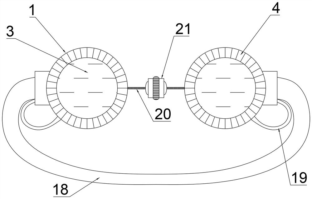
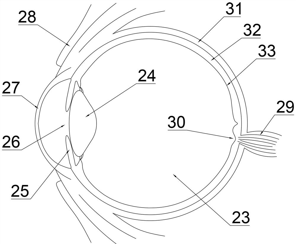
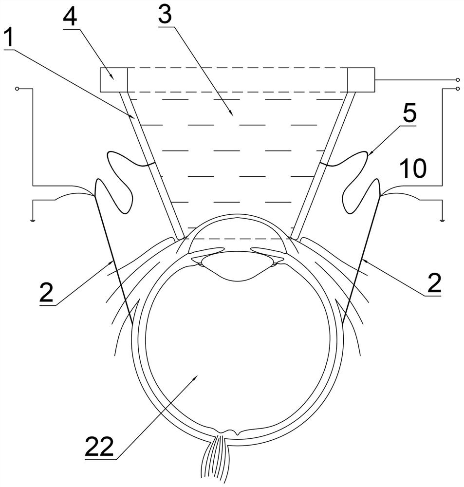
[0033] A microneedle device for retinal veins, such as Figure 1-6 As shown, it includes an iontophoresis chamber 1 and a microneedle 2. The iontophoresis chamber 1 is a hollow truncated cone with two openings inside and contains a therapeutic medium 3. One end of the iontophoresis chamber 1 is opened as a drug delivery end. , the administration end is buckled on the patient's eyeball 22, the cornea 27 and part of the conjunctiva 28 covering the eyeball 22, the other end opening of the iontophoresis chamber 1 is the observation end, and the observation end is provided with the first electrode 4, the iontophoresis chamber The side wall of 1 is evenly opened and connected to insulating hoses 5 respectively, and each insulating hose 5 is connected to a microneedle ...


PUM


Abstract

A retinal vein microneedle device belongs to a medical device, comprising an iontophoresis chamber and a microneedle, wherein the iontophoresis chamber is a hollow structure with openings at both ends and contains a therapeutic medium, and one end of the iontophoresis chamber is opened as The drug delivery end is buckled on the eyeball of the patient, and the other end is opened as the observation end, and the observation end is provided with a first electrode, the side wall of the iontophoresis chamber is evenly opened and connected to insulating hoses respectively, each insulating soft tube. The tubes are respectively connected with a microneedle; the microneedle is a hollow tubular shape, the front end of the microneedle is a second electrode, and the tail end of the microneedle is connected with an insulating hose, and under the guidance of the second electrode, the iontophoresis room medicine The ions are introduced into the mid-posterior segment of the patient's eyeball, with the first electrode and the second electrode having opposite polarities. The invention has the advantages of large medicine delivery range, high medicine delivery efficiency, small side reaction to patients and side injury of operation, and good operation effect.

Description

technical field [0001] The invention belongs to the technical field of medical devices, in particular to a microneedle device for retinal vein medication. Background technique [0002] For the treatment of fundus diseases, people need to apply medicines to the eyes, and various means have been used in order to treat diseases of the posterior segment of the eyes with medicines. Eye drops are the most common ophthalmic preparations. Eye drops are dripped into the conjunctival sac, first combined with tears, and transported into the eye through the cornea. The drug loss is serious, with less than 5% of the drug administered. Can reach the eye, long diffusion distance and convection of the aqueous humor, the drug can hardly reach the posterior segment of the eye after entering the eye; systemic administration is another way to treat eye diseases, but due to the aqueous humor barrier and blood - The isolation of the retinal barrier makes it difficult for drugs to enter the eyeba...

Claims


Application Information

Patent Timeline
Patent Type & Authority Patents(China)
IPC IPC(8): A61F9/00A61M37/00A61N1/30
CPCA61F9/0017A61M37/0015A61N1/306
Inventor 陶玥张自平王雪
Owner 郑州医笃筑工智能科技有限公司
Why Eureka
Try Eureka