Electron microscope quantitative detection method for virus particles by taking nanoparticles as reference substance

A quantitative detection method and virus particle technology, applied in the field of electron microscopy quantitative detection of virus particles, can solve problems such as errors, and achieve the effect of eliminating false negatives and increasing correctness

Pending Publication Date: 2020-06-09
智享生物(苏州)有限公司
View PDF5 Cites 4 Cited by
  • Summary
  • Abstract
  • Description
  • Claims
  • Application Information

AI Technical Summary

Problems solved by technology

Although the above-mentioned traditional electron microscope virus quantification method is very intuitive and has been used for more than half a century, there will be some errors because the recovery rate of the virus is not necessarily 100% during ultra-high speed centrifugation.

Method used


Image

  • Electron microscope quantitative detection method for virus particles by taking nanoparticles as reference substance
  • Electron microscope quantitative detection method for virus particles by taking nanoparticles as reference substance
  • Electron microscope quantitative detection method for virus particles by taking nanoparticles as reference substance

Examples


Embodiment 1

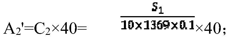
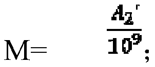
[0023] A method for quantitative detection of virus particles using nanoparticles as a reference substance, comprising the following steps:

[0024] (1) Mix 20ml of cell harvesting solution containing virus particles with 20ml of 1×10 9 A DMEM medium with a diameter of 20nm nanoparticles was mixed; then ultra-high-speed centrifugation was performed under a centrifugal force of 11000×g to collect virus particles; after ultra-high-speed centrifugation, the supernatant of the mobile phone was filtered with a filter membrane with a pore size of 0.45 μm, Perform ultra-high-speed centrifugation again under a centrifugal force of 100,000×g, and combine the virus particles collected by two ultra-high-speed centrifuges;

[0025] (2) Resuspend the collected virus particles in 20 μL DMEM medium and transfer to a PCR tube, then add an equal volume of 4% agar; place it at room temperature for 30 minutes, and wait for it to coagulate into a block, use glutaraldehyde, polysaccharide Treat w...

Embodiment 2

[0029] A method for quantitative detection of virus particles using nanoparticles as a reference substance, comprising the following steps:

[0030] (1) Mix 20ml of cell harvesting solution containing virus particles with 20ml of 1×10 9 A DMEM medium with a diameter of 20nm nanoparticles was mixed; then ultra-high-speed centrifugation was performed under a centrifugal force of 11000×g to collect virus particles; after ultra-high-speed centrifugation, the supernatant of the mobile phone was filtered with a filter membrane with a pore size of 0.45 μm, Perform ultra-high-speed centrifugation again under a centrifugal force of 100,000×g, and combine the virus particles collected by two ultra-high-speed centrifuges;

[0031] (2) Resuspend the collected virus particles in 20 μL DMEM medium and transfer to a PCR tube, then add an equal volume of 4% agar; place it at room temperature for 30 minutes, and wait for it to coagulate into a block, use glutaraldehyde, polysaccharide Treat w...

Embodiment 3

[0035] A method for quantitative detection of virus particles using nanoparticles as a reference substance, comprising the following steps:

[0036] (1) Mix 20ml of cell harvesting solution containing virus particles with 20ml of 1×10 9 A DMEM medium with a diameter of 20nm nanoparticles was mixed; then ultra-high-speed centrifugation was performed under a centrifugal force of 11000×g to collect virus particles; after ultra-high-speed centrifugation, the supernatant of the mobile phone was filtered with a filter membrane with a pore size of 0.45 μm, Perform ultra-high-speed centrifugation again under a centrifugal force of 100,000×g, and combine the virus particles collected by two ultra-high-speed centrifuges;

[0037] (2) Resuspend the collected virus particles in 20 μL DMEM medium and transfer to a PCR tube, then add an equal volume of 4% agar; place it at room temperature for 30 minutes, and wait for it to coagulate into a block, use glutaraldehyde, polysaccharide Treat w...


PUM

PropertyMeasurementUnit
diameteraaaaaaaaaa
diameteraaaaaaaaaa

Abstract

The invention discloses an electron microscope quantitative detection method for virus particles by taking nanoparticles as a reference substance. The method comprises the following steps: mixing a cell harvesting liquid containing the virus particles with a DMEM culture medium containing the nanoparticles with the diameter of 20-200nm; then carrying out ultra-high-speed centrifugal treatment, andcollecting the virus particles; re-suspending the collected virus particles into the DMEM culture medium, and then adding 3-5% of agar with the same volume as the DMEM culture medium; after the sample is coagulated into blocks, dehydrating and embedding the blocks, finally cutting the blocks into ultrathin slices with the size of 0.1 mu m as a sample, observing the number of the virus particles and the number of the nanoparticles with the area of 1369 mu m < 2 > under an electron microscope, and calculating the concentration of the nanoparticles in the electron microscope sample according tothe observed number of the virus particles and the number of the nanoparticles; and further calculating the concentration of the nanoparticles in the reference substance, and finally calculating the concentration of the virus particles in cell harvesting liquid. According to the method, errors existing in a traditional detection method can be effectively avoided.

Description

Technical field: [0001] The invention relates to the field of biotechnology, in particular to an electron microscope quantitative detection method for virus particles using nanoparticles as a reference substance. Background technique: [0002] The production of biomedical products is different from traditional chemical drugs. Most of them are produced by organisms such as cells and bacteria, or extracted from biological fluids. Therefore, many microorganisms and viruses that are not found in chemical preparations are hidden in the production process. Or the possibility of contamination by substances such as host nucleic acid and protein, which will cause certain harm to healthy users when the drug is used. In order to ensure the quality and safety of protein drugs, producers must check the sources of upstream materials, intermediate products in the manufacturing process, and final products to confirm their safety for users. [0003] Biosafety testing items are generally div...

Claims


Application Information

Patent Timeline
Patent Type & Authority Applications(China)
IPC IPC(8): G01N23/2251G01N23/2202
CPCG01N23/2251G01N23/2202G01N2223/07G01N2223/102G01N2223/612G01N2223/635
Inventor 李智李育修
Owner 智享生物(苏州)有限公司
Why Eureka
Try Eureka