Method for enhancing contrast of microscopic CT plant sample

A contrast and plant technology, applied in the biological field, can solve problems such as difficult structure edges, few researches, and small image contrast

Active Publication Date: 2020-07-10
INST OF BOTANY CHINESE ACAD OF SCI
View PDF4 Cites 1 Cited by
  • Summary
  • Abstract
  • Description
  • Claims
  • Application Information

AI Technical Summary

Problems solved by technology

In the late 1990s, CT began to be used in plant research to study root structure, morphological development, and plant fossils, but it is rarely found in softer tissues such as young organs, tissues with low or uniform density. Research
These lead to no significant difference in ray transmittance, and it is very difficult to distinguish the edges of sample structures with similar densities. The reconstructed image has very little contrast and high noise (Rousseau et al., 2015)

Method used


Image

  • Method for enhancing contrast of microscopic CT plant sample
  • Method for enhancing contrast of microscopic CT plant sample
  • Method for enhancing contrast of microscopic CT plant sample

Examples


Embodiment Construction

[0034] The experimental methods used in the following examples are conventional methods unless otherwise specified.

[0035] The materials and reagents used in the following examples can be obtained from commercial sources unless otherwise specified.

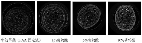
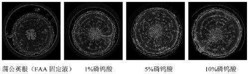
[0036] Dissolve phosphotungstic acid in FAA fixative solution to obtain a phosphotungstic acid solution with a mass concentration of 1 to 10%, wherein the composition of FAA fixative solution is as follows: FAA fixative solution (100ml): 90ml of 50% ethanol aqueous solution, 5ml of glacial acetic acid , formaldehyde 5ml.

[0037] The following examples were subjected to carbon dioxide critical point drying in a critical point dryer (CPD300, Leica).

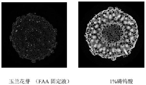
[0038] 1. Magnolia flower buds were put into FAA fixative solution and 1% phosphotungstic acid solution respectively, turned upside down several times to remove air bubbles, until the sample sank into the solution, dyed and fixed at 4°C for 2 days, and then used 70%, 80%, 90% and ...


PUM


Abstract

The invention discloses a method for enhancing contrast of a microscopic CT plant sample. The method comprises the following steps: 1) dissolving phosphotungstic acid in an FAA stationary liquid to obtain a phosphotungstic acid solution; 2) soaking a plant part to be detected in the phosphotungstic acid solution for fixing and dyeing treatment; and (3) sequentially carrying out dehydration and carbon dioxide critical point drying on the plant part fixed and dyed in the step (2) so as to enhance the contrast ratio of the microscopic CT of the plant sample. Microscopic CT scanning (Skyscan1172,Brook Company) is performed on the sample subjected to carbon dioxide critical point drying, the scanning voltage can be 41V-59V, the resolution can be 0.56-1.76 mu m, a cross section picture is obtained through NRecon software reconstruction after the sample is scanned, the contrast ratio of plant samples at different parts is greatly improved, and the contrast ratio of parts rich in cytoplasm and protein, such as magnolia flower buds, is more remarkably improved.

Description

technical field [0001] The invention relates to a method for enhancing the contrast of micro-CT plant samples, which belongs to the field of biotechnology. Background technique [0002] Micro-CT technology has been born for more than 40 years, and it has played a huge role in medicine, and has many applications in geology and materials science. In the late 1990s, CT began to be used in plant research to study root structure, morphological development, and plant fossils, but it is rarely found in softer tissues such as young organs, tissues with low or uniform density. Research. [0003] Micro-CT imaging requires X-rays, while X-ray contrast imaging relies on the density, thickness, and atomic number of the sample (Lusic and Grinstaff, 2013). Since plant tissues are mainly composed of light elements, the X-ray absorption rate is low (about 20-40%), and the imaging effect of samples with an absorption rate of about 65-90% is the best. In addition, young plant tissues have n...

Claims


Application Information

Patent Timeline
Patent Type & Authority Applications(China)
IPC IPC(8): G01N1/30G01N23/046
CPCG01N1/30G01N23/046
Inventor 徐秀苹冯旻梁荣花靳婉青
Owner INST OF BOTANY CHINESE ACAD OF SCI
Why Eureka
Try Eureka