Ultrasound contrast video data analysis method based on composite neural network

A technology of neural network and contrast-enhanced ultrasound, which is applied in image analysis, image data processing, instruments, etc., to achieve rapid analysis and reduce the demand for computer computing power

Active Publication Date: 2020-07-10
THE FIRST AFFILIATED HOSPITAL OF SUN YAT SEN UNIV
View PDF4 Cites 3 Cited by
  • Summary
  • Abstract
  • Description
  • Claims
  • Application Information

AI Technical Summary

Problems solved by technology

[0005] However, video is composed of continuous single-frame images. A 1-2 minute CEUS video contains thousands of frames of images, and the data size often reaches hundreds of megabytes. Therefore, powerful computer computing power is required to analyze it.

Method used


Image

  • Ultrasound contrast video data analysis method based on composite neural network
  • Ultrasound contrast video data analysis method based on composite neural network
  • Ultrasound contrast video data analysis method based on composite neural network

Examples


Embodiment 1

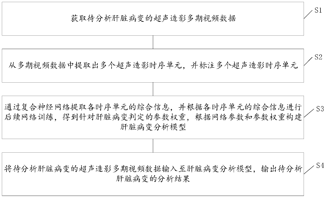
[0030] See figure 1 , an embodiment of the present invention provides a composite neural network-based method for analyzing video data of contrast-enhanced ultrasound, including steps S1-S4;

[0031] S1. Acquiring multi-phase contrast-enhanced ultrasound video data of liver lesions to be analyzed.

[0032] In this embodiment, step S1 specifically includes: collecting multi-stage segmented video data of the liver lesion to be analyzed to obtain multi-stage video data;

[0033] Among them, each phase includes arterial phase, portal phase and delay phase.

[0034] As a preferred embodiment of the present invention, after step S1, it further includes: clipping the multi-phase video data, and removing redundant information to retain the target lesion and a certain range of liver parenchyma around the lesion.

[0035] S2. Extract a plurality of time-sequence units of contrast-enhanced ultrasound from the multi-phase video data, and mark the time-series units of contrast-enhanced u...

Embodiment 2

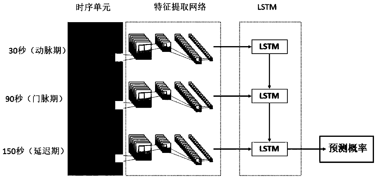
[0045] As an optional embodiment, based on the CEUS diagnosis of hepatocellular carcinoma lesions, the fourth generation convolutional neural network ResNet and the long short-term memory network (Long Short-Term Memory, LSTM) are used to form a composite network to realize CEUS time series Extraction and utilization of information;

[0046] Such as figure 2 As shown, multiple CEUS videos of this case were acquired, including the arterial phase from 10 seconds to 30 seconds, the portal vein phase from 31 seconds to 120 seconds, and the delay period from 120 seconds to 360 seconds, which are video data collected and stored in segments; from the video At 20 seconds, 90 seconds, 150 seconds and several seconds before and after, select single-frame images that clearly show the target lesion, and select 15 frames at each time point; one frame at each time point constitutes a time-series unit containing 3 frames of images; Extract 15 time-series units; crop each frame of image to ...


PUM


Abstract

The invention discloses an ultrasound contrast video data analysis method based on a composite neural network. The method comprises the following steps: acquiring ultrasound contrast multi-stage videodata of a to-be-analyzed liver lesion; extracting a plurality of ultrasonic contrast time sequence units from the multi-phase video data; marking a plurality of ultrasonic contrast time sequence units; extracting comprehensive information of each time sequence unit through a composite neural network; performing subsequent network training according to the comprehensive information of each time sequence unit; obtaining a parameter weight for liver lesion judgment, constructing a liver lesion analysis model according to the network parameter and the parameter weight, finally inputting ultrasound contrast multi-stage video data of a to-be-analyzed liver lesion into the liver lesion analysis model, and outputting the analysis result of the to-be-analyzed liver lesion. By adopting the embodiment provided by the invention, the CEUS time sequence information can be fully utilized, and the computer computing power requirement for video analysis is reduced, so that the liver lesion to be analyzed can be quickly analyzed.

Description

technical field [0001] The invention relates to the technical fields of medical treatment and data processing, in particular to a method for analyzing video data of contrast-enhanced ultrasound based on a composite neural network. Background technique [0002] CEUS is one of the three conventional imaging methods for evaluating liver lesions (the other two are CT and MR), and its wide application has accumulated a large amount of valuable data for clinical research. However, due to the heterogeneity of data caused by different instruments, different imaging conditions, and different operators, the development of computer-aided diagnosis technology for CEUS is less, which greatly limits its technical development. [0003] CEUS video can provide image information closely related to the lesion from blood perfusion, reflecting a continuous and dynamic enhancement-regression process, that is, sequential information. This is the biggest advantage of CEUS over other imaging method...

Claims


Application Information

Patent Timeline
Patent Type & Authority Applications(China)
IPC IPC(8): G06T7/00
CPCG06T7/0012G06T2207/10132G06T2207/20081G06T2207/30056G06T2207/30096Y02A90/10
Inventor 胡航通王伟陈立达阮思敏匡铭谢晓燕吕明德
Owner THE FIRST AFFILIATED HOSPITAL OF SUN YAT SEN UNIV
Why Eureka
Try Eureka