Electrocardiosignal classification method and device, electronic equipment and storage medium

A technology of electrocardiographic signal and classification method, applied in character and pattern recognition, instrument, biological neural network model, etc., can solve problems such as huge labeling cost and huge size

Pending Publication Date: 2020-08-04
GUANGZHOU SHIYUAN ELECTRONICS CO LTD
View PDF7 Cites 5 Cited by
  • Summary
  • Abstract
  • Description
  • Claims
  • Application Information

AI Technical Summary

Problems solved by technology

However, during continuous ECG monitoring, the ECG is recorded for at least 24 hours and contains 100,000 to 200,000 heartbeat waveforms, a number that is usually too large to be individually labeled by a cardiologist
[0003] Automatic classification is an essential function in ECG signal monitoring. In clinical practice, various types of features ba

Method used


Image

  • Electrocardiosignal classification method and device, electronic equipment and storage medium
  • Electrocardiosignal classification method and device, electronic equipment and storage medium
  • Electrocardiosignal classification method and device, electronic equipment and storage medium

Examples


Embodiment 1

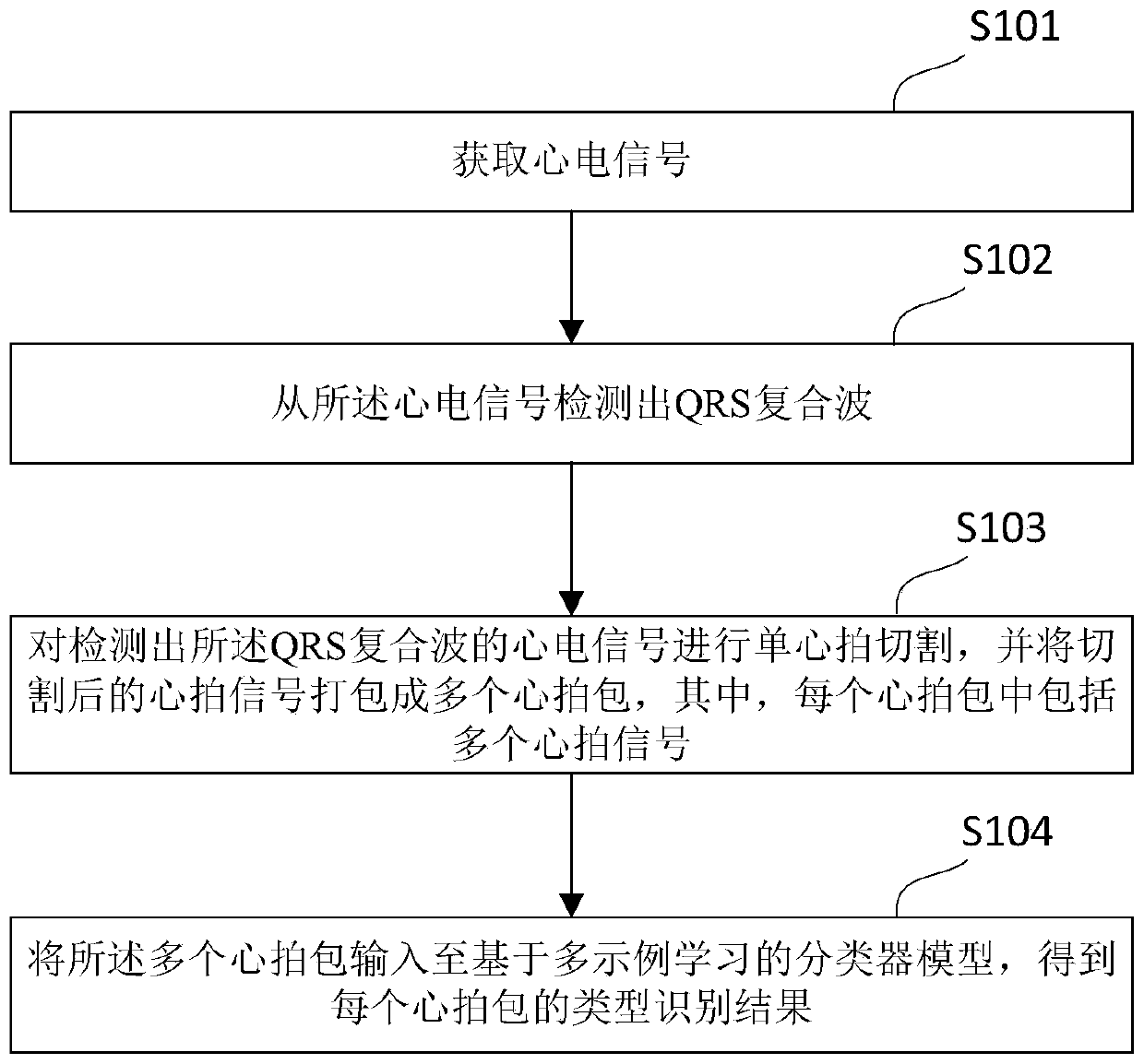
[0092] figure 1 A flowchart of a method for classifying ECG signals provided in Embodiment 1, according to figure 1 As shown, the ECG signal classification method in Embodiment 1 includes the following steps:

[0093] Step S101: Acquire ECG signals.

[0094] The electrocardiographic signal is the signal of the electrical activity change generated by the heart during each cardiac cycle recorded from the body surface, and is usually recorded graphically through an electrocardiogram (ECG). In the embodiment of the present application, multi-channel synchronous data may be used to collect and store human heart signals, background noise, and electrocardiographic signals. For example, electrocardiographic signals can be collected through electrocardiographic leads and sensors, and analog signals of human physiological parameters can be converted into digital signals by analog-to-digital converters, which are stored in memory.

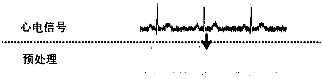
[0095] like figure 2 As shown, in an example, afte...

Embodiment 2

[0123] Figure 5 A flowchart of a method for classifying electrocardiographic signals provided in Embodiment 2, according to Figure 5 As shown, the electrocardiographic signal classification method in the second embodiment includes the following steps:

[0124] Step S501: Obtain ECG signals.

[0125] Step S502: Detecting the QRS complex wave from the electrocardiographic signal.

[0126] Specifically, such as Image 6 As shown, the described detection of the QRS complex from the electrocardiographic signal may include the following sub-steps:

[0127] Step S5021: Perform discrete wavelet decomposition on the ECG signal to obtain a preset number of wavelet decomposition coefficients.

[0128] In an example, Daubechies4 (Db4) wavelet may be used to perform discrete wavelet decomposition on the preprocessed ECG signal. The wavelet decomposition coefficients are determined by the number of layers of wavelet decomposition. In a preferred example, the number of wavelet decompo...

Embodiment 3

[0172] Figure 9 A flow chart of a method for classifying electrocardiographic signals provided in Embodiment 3, according to Figure 9 As shown, the electrocardiographic signal classification method in the third embodiment includes the following steps:

[0173] Step S901: Acquiring ECG signals.

[0174] Step S902: Detecting the QRS complex wave from the electrocardiographic signal.

[0175] Step S903: Cutting the electrocardiographic signal from which the QRS complex wave is detected into a single heart beat, and packaging the cut heart beat signal.

[0176] Step S904: Input the plurality of cardiac beat packets into the second deep network model;

[0177] Step S905: Using the second deep network model to extract features of each heartbeat in the plurality of heartbeat packets, and input the extracted features to the support vector machine.

[0178] Step S906: Using the support vector machine to classify the features of each heartbeat, and obtain the type identification r...


PUM


Abstract

The embodiment of the invention relates to an electrocardiosignal classification method and device, electronic equipment and a storage medium. The electrocardiosignal classification method disclosed by the embodiment of the invention comprises the following steps: acquiring electrocardiosignals; detecting a QRS complex wave from the electrocardiosignal; carrying out single heart beat cutting on the electrocardiosignals with the detected QRS composite waves and then packaging the electrocardiosignals into a plurality of heart beat packets; inputting the plurality of heart beat packets into a classifier model based on multi-example learning, obtaining a type identification result of each heart beat packet, wherein when the classifier model based on multi-example learning is used for classification, each heart beat packet is used as an example packet, each heart beat signal in the heart beat packet is used as an example in the example packet, and the type identification result comprises anormal rhythm type and an abnormal rhythm type. The electrocardiosignal classification method provided by the embodiment of the invention effectively reduces the time and labor cost spent in performing machine learning classification by manually labeling the heart beat.

Description

technical field [0001] The embodiments of the present application relate to the technical field of electrocardiographic signal classification, and in particular, relate to a method, device, electronic device, and storage medium for electrocardiographic signal classification. Background technique [0002] Heart disease is a common cardiology disease caused by structural damage or abnormal function of the heart. Heart disease can be detected by electrocardiogram (ECG) signal, which is a waveform record of bioelectrical changes produced by the myocardium. Occasional abnormalities in the heart can be monitored through a 24-hour dynamic electrocardiogram, which can more accurately determine whether there is a heart disease. Conventional ECG signals require annotated heartbeats labeled by trained cardiologists for classification. However, during continuous ECG monitoring, the ECG recording lasts at least 24 hours and contains 100,000-200,000 beat waveforms, which is usually too l...

Claims


Application Information

Patent Timeline
IPC IPC(8): G06K9/00G06K9/62G06N3/04
CPCG06N3/044G06N3/045G06F2218/08G06F2218/12G06F18/2411
Inventor 胡静
Owner GUANGZHOU SHIYUAN ELECTRONICS CO LTD
Why Eureka
Try Eureka