Method for detecting circulating miRNA by using DNA machine based on quantum dot micelle spherical nucleic acid

A technology of quantum dots and DNA enzymes, applied in the field of biomedicine, can solve the problems of inconvenient functional nucleic acid integration, small particle size of water-soluble quantum dots, etc., and achieve the effect of high sensitivity and multivariate detection.

Active Publication Date: 2020-08-28
SOUTHEAST UNIV
View PDF1 Cites 1 Cited by
  • Summary
  • Abstract
  • Description
  • Claims
  • Application Information

AI Technical Summary

Problems solved by technology

More importantly, the particle size of water-soluble quantum dots prepared by the ligand exchange

Method used


Image

  • Method for detecting circulating miRNA by using DNA machine based on quantum dot micelle spherical nucleic acid
  • Method for detecting circulating miRNA by using DNA machine based on quantum dot micelle spherical nucleic acid
  • Method for detecting circulating miRNA by using DNA machine based on quantum dot micelle spherical nucleic acid

Examples


Embodiment 1

[0024] Sequence design of three targets, three DNases, and three substrates

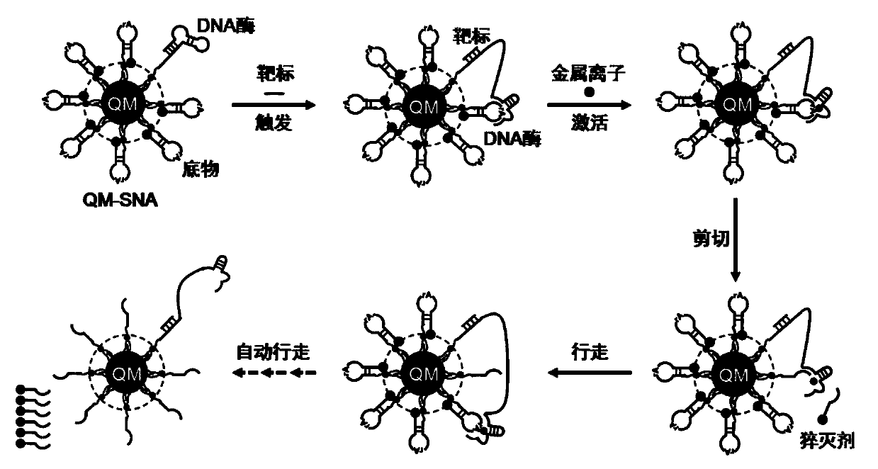
[0025] Such as figure 1 As shown, the corresponding DNA enzymes (DNAzyme 1-3) and substrates (Substrate 1-3 ). The DNase sequence mainly includes three parts: the target binding region, the (substrate) track binding region, the catalytic center region, and the modified amino group at the 5' end for coupling to the QM surface. Among them, the target binding region sequence is the complementary sequence of the target, which is used to hybridize with the target to open the stem-loop structure of DNase; the orbital binding region includes two sequences of the upper and lower arms, which are used to bind to the substrate; the catalytic center region is used to bind metal ions Recognizes and cuts the rA site. The substrate sequence mainly includes the complementary sequence of the upper and lower arms, the rA site, the modified amino group at the 3' end is used for coupling to the QM surface, and the BH...

Embodiment 2

[0029] Preparation of three-color QM-SNA loaded with DNase and substrate

[0030] The three-color QM-SNA is prepared by chemical cross-linking, and the preparation process is the same. Take 50 μL of QM solution (concentration: 50 nM), 10 μL of EDC solution (100 μM) and Sulfo-NHS solution (50 μM) into 0.01M, pH7.4 phosphate buffer solution (total volume 500 μL), mix and stir for 15 min Afterwards, 25 μL of DNase solution (45 nM) and 225 μL of substrate nucleic acid solution (455 nM) were sequentially added, reacted for 4 hours at an appropriate stirring speed, and purified by ultrafiltration.

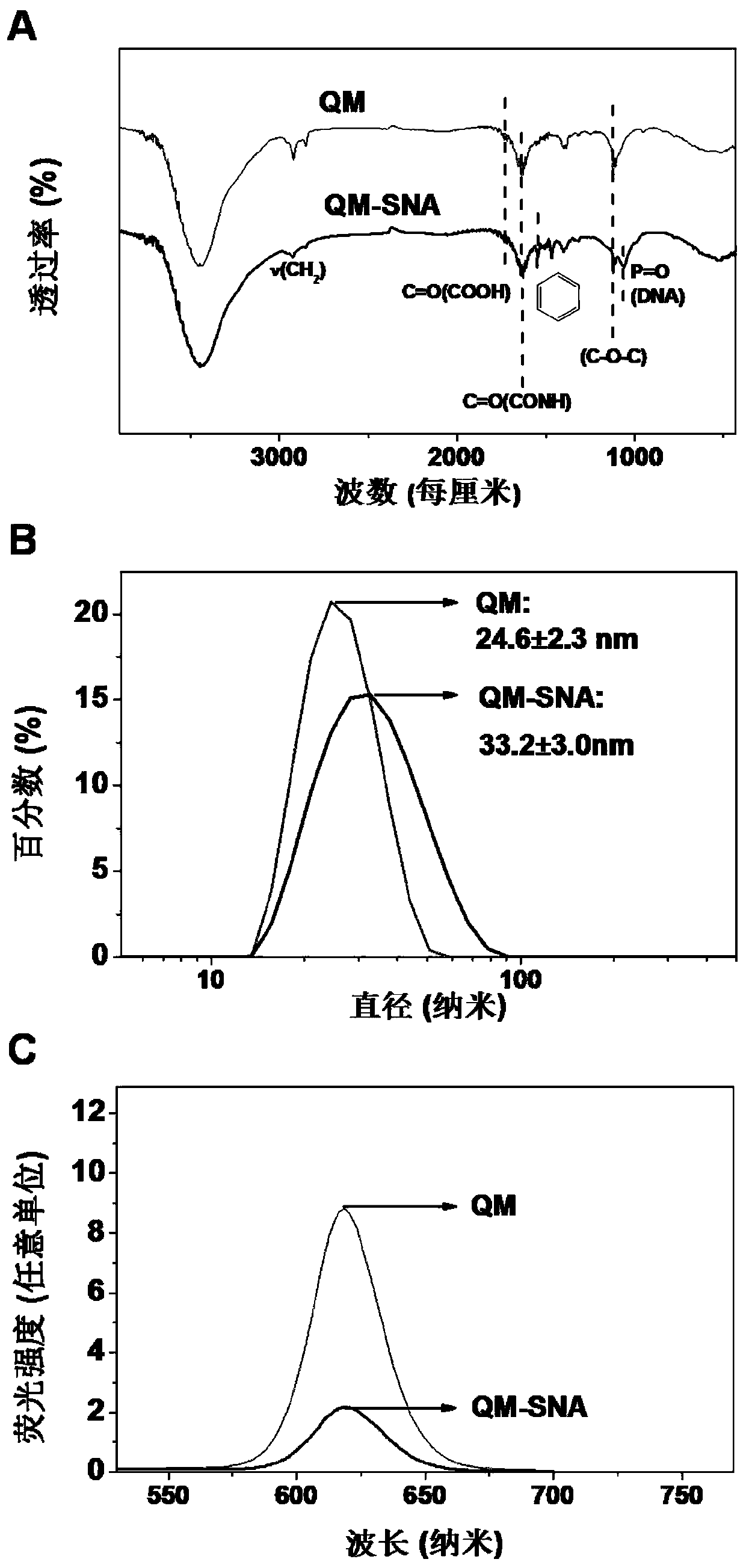
[0031] Characterize the composition, hydrodynamic size, fluorescence and other properties of QM-SNA, the results are as follows figure 2 shown. figure 2 A, Compared with QM, the infrared spectrum of QM-SNA appeared the characteristic peak of CONH bond attributed to the surface coupling (1635cm -1 ), attributed to the P=O bond characteristic peak of nucleotide composition (1062cm -1...

Embodiment 3

[0033] QM surface target-triggered DNase walking

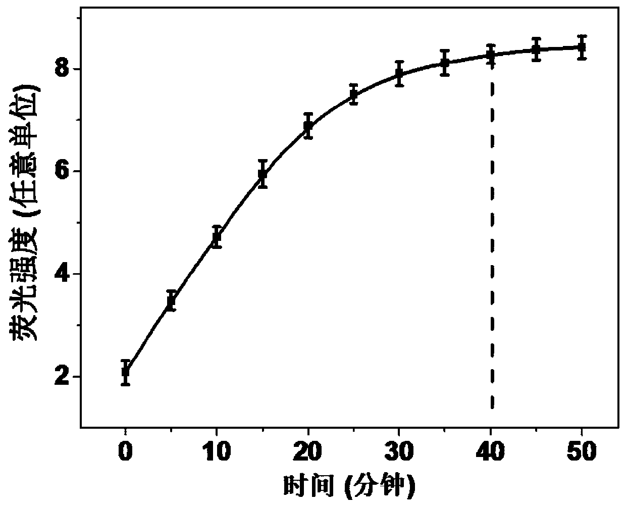
[0034] Add QM-SNA (10nM) and target miRNA (50pM) solution to Tris-HCl (25mM, pH 7.4) buffer solution sequentially, with a total volume of 195μL, and fully react at 37°C for 1h to fully hybridize the target miRNA. Then add 5 μL of Zn 2+ (200mM) solution, measure the fluorescence spectrum every 5min.

[0035] The fluorescence intensity of QM changes as image 3 Shown, Zn 2+ With the extension of time after the addition, the fluorescence of QM gradually increased, and the fluorescence signal reached the maximum at about 40 min, which indicated the continuous walking process of DNase on the surface of QM.


PUM


Abstract

The invention discloses a method for detecting circulating miRNA by using a DNA machine based on quantum dot micelle spherical nucleic acid. The specific implementation method comprises the followingthree parts: (1) preparing multicolor quantum dot micelle spherical nucleic acid (QM-SNA) which is loaded with a DNA enzyme sequence and a quenching agent-modified substrate sequence; (2) triggering atarget so as to achieve automatic walking of DNA enzymes along a DNA track (the substrate sequence) on the surface of QM under the assistance of metal ions; and (3) measuring fluorescence signals ofthe multicolor QM through adoption of a fluorospectro photometer after completion of walking so as to realize detection and analysis of miRNA. According to the method for detecting the circulating miRNA by using the DNA machine based on the multicolor QM-SNA, a DNA enzyme walker is used for mediating amplification of the QM fluorescence signals, so that high-sensitivity detection of the miRNA is realized; and multi-element detection of miRNA is realized through adoption of the multi-color QM-SNA, and a new method is provided for accurate detection of the circulating miRNA.

Description

technical field [0001] The invention relates to a method for detecting circulating miRNA using a DNA machine based on quantum dot micellar spherical nucleic acid, and belongs to the technical field of biomedicine. Background technique [0002] microRNA (miRNA) is a class of endogenous non-coding RNA with a length of about 22 bases. It can regulate the expression level of post-transcriptional genes by inhibiting the expression of targeted mRNA. At the same time, miRNA can regulate cell development and cell differentiation. , immune response and other important physiological processes can also be used as proto-oncogenes or tumor suppressor genes in different cancer cells to regulate the abnormal expression of corresponding pathological processes. A number of studies have shown that the abnormal expression of circulating miRNAs that are stable in blood and body fluids is closely related to the occurrence and development of various cancers, and has become an important class of t...

Claims


Application Information

Patent Timeline
IPC IPC(8): C12Q1/6816
CPCC12Q1/6816C12Q2525/207C12Q2521/345C12Q2563/107C12Q2563/137
Inventor 孙清江
Owner SOUTHEAST UNIV
Why Eureka
Try Eureka