Application of oncolytic virus in treatment on uveal melanoma, marker of treatment effect and detection reagent thereof

A technology for melanoma and melanoma cells, applied in the field of kits containing the marker or detection preparation, can solve the problems of patient death, no observed mortality, etc.

Active Publication Date: 2020-10-30
BEIJING NEUROSURGICAL INST
View PDF3 Cites 3 Cited by
  • Summary
  • Abstract
  • Description
  • Claims
  • Application Information

AI Technical Summary

Problems solved by technology

With limited therapies available to treat this chemoresistant tumor, metastases occur within 15 years and cause death in approximately 90% of patients [6]
However, no difference in mortality was observed between conservative and surgical management

Method used


Image

  • Application of oncolytic virus in treatment on uveal melanoma, marker of treatment effect and detection reagent thereof
  • Application of oncolytic virus in treatment on uveal melanoma, marker of treatment effect and detection reagent thereof
  • Application of oncolytic virus in treatment on uveal melanoma, marker of treatment effect and detection reagent thereof

Examples


Embodiment 1

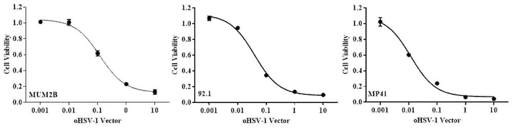
[0118] Example 1. Sensitivity test of human UM cell line to oHSV-1 backbone.

[0119] The human UM cell lines MUM2B, 92.1 and MP41 described in Section 1 of Materials and Methods were used as models to test the sensitivity of oHSV-1 to UM, respectively. These three cell lines were used to represent the major genotypes of UM. Sensitivity of oHSV-1 vectors to UM cells was tested using the cell survival assay described in Section 3 of Materials and Methods ( figure 2 ). The results showed that UM cells were sensitive to oHSV-1 in a dose-dependent manner. IC for each of the three cell lines 50 They are 0.1211MOI, 0.03897MOI and 0.01195MOI, respectively. These results suggest that UM cells are sensitive to the genetically engineered oHSV-1 virus.

[0120] 24 hours after cell seeding, medium was replaced with diluted oHSV-1 added to complete medium for MUM2B and MP41 cells in DMEM with 10% fetal bovine serum for 92.1 cell line , using 10% fetal bovine serum in RPMI-1640 mediu...

Embodiment 2

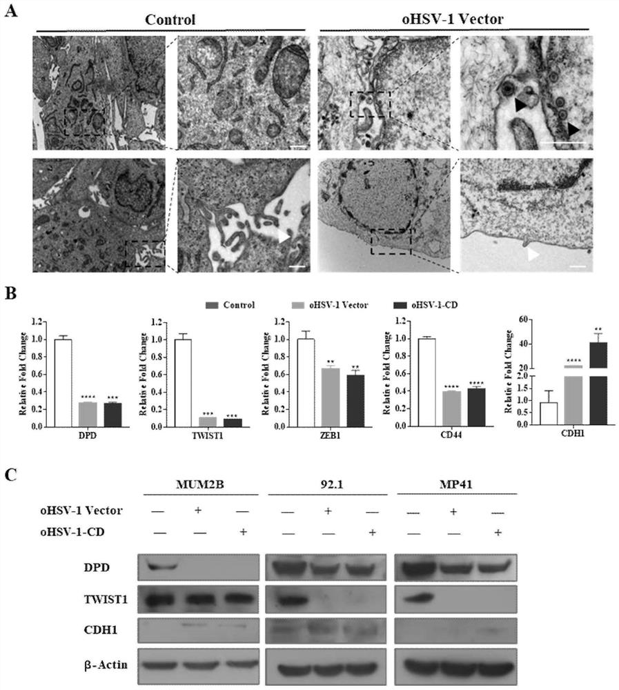
[0121] Example 2. Test of the effect of oncolytic HSV-1 on MUM2B cell phenotype and gene expression

[0122]To further test the tumor-killing effectiveness of oHSV-1, according to the method in Section 5 of Materials and Methods, the ultramicroscopic morphologies of MUM2B cells after oHSV-1 infection (infection dose MOI of 0.1, infection time 72 h) were observed by electron microscope TEM. structure( image 3 A). The virus was observed in the nucleus and cytoplasm (black arrows). Microvilli were reduced in infected cells compared to uninfected cells (white arrows). These data suggest that the EMT (epithelial-mesenchymal transition) process is inhibited by oHSV-1 infection.

[0123] Next, with MUM2B cells as the control and oHSV-1 infection as the test group, the infection dose MOI is 0.1, the present invention further detects whether oHSV-1 infection can inhibit EMT. Using the primers shown in Table 2, the expression abundance of EMT marker genes was tested by conventional...

Embodiment 3

[0127] Example 3. Detection of the effectiveness of oHSV-1-CD / 5-FC on UM cell line

[0128] Using the oHSV-1-CD constructed by the methods of Section 2 of Materials and Methods, in vitro experiments were performed against the oHSV-1-CD / 5-FC model to determine whether the recombinant virus functions in the UM cell line. The efficacy of oHSV-1-CD / 5-FC treatment was examined using three human UM cell lines, MUM2B, 92.1 and MP41. Seed 5,000 cells in each plate at 0MOI, 0.001MOI, 0.01MOI, 0.1MOI, 1MOI, 10MOI, oHSV-1-CD / 5 from left to right using the method in Section 10 of Materials and Methods The concentration of 5-FC in the -FC group was 50 μg / mL, and three rows were repeated. The grayscale drop within each treatment is shown by IVIS images. The decrease in IVIS gray scale was significantly greater in cells receiving combination treatment compared to the group treated with oHSV-1-CD alone ( Figure 4 A). ROIs were measured to quantify pixel intensities in IVIS images ( Fig...


PUM


Abstract

The invention discloses a preparation for treating, preventing and / or slowing down uveal melanoma, the preparation comprises oncolytic herpes simplex virus type 1 capable of expressing cytosine deaminase genes and 5-fluorocytosine, and the oncolytic herpes simplex virus type 1 and the 5-fluorocytosine are combined to significantly reduce the volume of the uveal melanoma and improve the lifetime ofa subject. The invention further discloses a marker for evaluating the treatment effect of uveal melanoma through the preparation, and the marker is selected from markers capable of representing theepithelial-mesenchymal transition degree and mainly comprises IL-6, DPD, TWIST1, ZEB1, CD44 and CDH1.

Description

technical field [0001] The invention belongs to the field of medicine, and relates to a marker for evaluating the therapeutic effect of uveal melanoma and a detection preparation thereof, in particular to a marker for evaluating the treatment of oncolytic herpes simplex virus containing cytosine deaminase gene combined with 5-FC A marker for the therapeutic effect of uveal melanoma, a detection preparation thereof, and a corresponding kit containing the marker or detection preparation. Background technique [0002] As an emerging and important tumor treatment method, oncolytic virus can selectively kill and destroy tumor cells. A variety of oncolytic RNA and DNA viruses that can selectively replicate and cause direct lysis of tumor cells have been widely reported [1,2] . Most clinical studies have focused on adenoviruses, poxviruses, herpes simplex virus type 1 (oHSV-1), measles, and reoviruses. [0003] Uveal melanoma (UM) is the most common primary intraocular malignanc...

Claims


Application Information

Patent Timeline
Patent Type & Authority Applications(China)
IPC IPC(8): C12Q1/6886G01N33/577G01N33/574A61K48/00A61K35/763A61K38/50A61K31/513A61P35/00
CPCC12Q1/6886G01N33/577G01N33/57484G01N33/57407A61K48/005A61K48/0008A61K35/763A61K38/50A61K31/513C12Y305/04001A61P35/00C12Q2600/106C12Q2600/158C12Q2600/166G01N2800/60A61K2300/00Y02A50/30
Inventor 刘福生刘思思张俊文
Owner BEIJING NEUROSURGICAL INST
Why Eureka
Try Eureka