Cancer cell identification and diagnosis system

A diagnostic system and cancer cell technology, applied in character and pattern recognition, image data processing, instruments, etc., can solve problems such as difficult recognition

Pending Publication Date: 2020-11-03
GUILIN UNIV OF ELECTRONIC TECH
View PDF0 Cites 5 Cited by
  • Summary
  • Abstract
  • Description
  • Claims
  • Application Information

AI Technical Summary

Problems solved by technology

Due to the complex background of cell images, diverse and overlapping cell shapes, etc., it is difficult to detect and identify by computer

Method used


Image

  • Cancer cell identification and diagnosis system
  • Cancer cell identification and diagnosis system
  • Cancer cell identification and diagnosis system

Examples


Embodiment

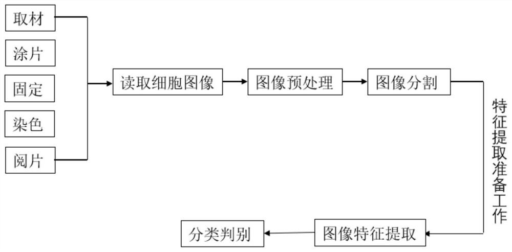
[0024] refer to figure 1 , a cancer cell identification and diagnosis system comprising,

[0025] Cell image preprocessing module:

[0026] First convert the cell picture into a digital image and process it in gray scale. In order to extract useful cell features in the image more accurately, it is necessary to remove small, discrete normal cells and additive noise in the image. In view of this situation, the improved open-close filter algorithm is used for image denoising, and the structural element B is selected to be twice the normal lymphocyte. For the noise smaller than the structure element B, use the opening operation to eliminate the pepper noise in the background, and then use the closing operation to eliminate the trachoma noise. For the noise larger than structural element B, the filtered image is binarized first, and the original image is superimposed and extracted. Image binarization is to convert the original image into a binary image, "0" represents the targe...


PUM


Abstract

The invention discloses a cancer cell recognition and diagnosis system, which comprises a cell image preprocessing module for selecting proper structural elements by adopting an improved mathematicalmorphology opening and closing filtering algorithm to remove background impurities and small normal discrete cells of a cell image; a cell image segmentation module used for extracting a cell nucleusregion by adopting an improved region growth algorithm, extracting a non-laminated cell body contour by adopting a watershed algorithm based on price tag control, and extracting a laminated cell contour by adopting a segmentation algorithm based on a snappe model; a cancer cell image feature extraction module used for calculating the nuclear-to-mass ratio of each region after segmentation, whetherthe cell nucleus size is uniform, whether nuclear staining is abnormal and whether the nucleus spacing is uniform. and a cell image classification and recognition module which recognizes cancer cellsthrough a neural network recognition technology. The system can be used for efficiently and accurately carrying out quantitative analysis and detection identification on cell images.

Description

technical field [0001] The invention relates to the technical field of cell image recognition, in particular to a cancer cell recognition and diagnosis system. Background technique [0002] Cancer is a common disease that endangers human health, and early diagnosis of cancer is the key to treatment. The use of cytological computer-aided diagnosis technology can effectively reduce the work intensity of doctors and improve the accuracy of diagnosis. Due to the complex background of cell images, diverse and overlapping cell shapes, it is difficult to detect and identify by computer. The invention makes new technological improvements to the typical medical image segmentation algorithm, and provides improved algorithms for non-overlapping cell images and overlapping cell images respectively. Contents of the invention [0003] The purpose of the present invention is to provide a cancer cell identification and diagnosis system for the deficiencies in the prior art. This diagno...

Claims


Application Information

Patent Timeline
Patent Type & Authority Applications(China)
IPC IPC(8): G06T7/11G06T7/13G06T7/136G06T7/187G06T7/00G06T5/00G06K9/62G06N3/04G06N3/08
CPCG06T7/11G06T7/13G06T7/136G06T7/187G06T7/0012G06T7/62G06T5/002G06T2207/10056G06T2207/20024G06T2207/20081G06T2207/20084G06T2207/30024G06N3/084G06N3/045G06F18/241
Inventor 车俐韩梦玲
Owner GUILIN UNIV OF ELECTRONIC TECH
Why Eureka
Try Eureka