Kit for quantitatively detecting lymphocyte subsets and detection method thereof

A lymphocyte and quantitative detection technology, applied in the field of immunological detection, can solve the problems of high reagent cost, difficult to meet the general application of detection technology, uneven hardware configuration of instruments, etc., to shorten the detection time, improve the scope of application and simple operation Effect

Active Publication Date: 2020-12-18
UB BIOTECHNOLOGY ZHEJIANG CO LTD
View PDF7 Cites 1 Cited by
  • Summary
  • Abstract
  • Description
  • Claims
  • Application Information

AI Technical Summary

Problems solved by technology

Most of the lymphocyte subset detection kits currently used in clinical practice are developed by the American BD company. This kit contains 7 kinds of monoclonal antibodies, which are labeled with 6 different fluoresceins, so the hardware requirements for flow cytometry are very high. , must be equipped with at least two lasers of 488nm and

Method used


Image

  • Kit for quantitatively detecting lymphocyte subsets and detection method thereof
  • Kit for quantitatively detecting lymphocyte subsets and detection method thereof
  • Kit for quantitatively detecting lymphocyte subsets and detection method thereof

Examples


Embodiment 1

[0028] A kit for quantitative detection of lymphocyte subsets, comprising the following components: 20 μL of monoclonal antibody mixture, and 500 μL of erythrocyte lysate.

[0029] Wherein, the content of each component of the monoclonal antibody mixture is as follows:

[0030] ① CD3 FITC, 0.24μg;

[0031] ② CD56 FITC, 0.6μg;

[0032] ③ CD45 PerCP-Cy5.5, 0.0192μg;

[0033] ④ CD8 PE, 0.006μg;

[0034] ⑤ CD19 PE, 0.075μg;

[0035] ⑥ CD4 PE-Cyanine7, 0.075μg.

[0036] Red blood cell lysate, the mass proportion of each component is as follows:

[0037] ① The concentration of sodium citrate dihydrate is 1.764%;

[0038] ② The concentration of isobutanol is 0.888%;

[0039] ③ Formaldehyde concentration 5.256%;

[0040] ④ Sodium perchlorate concentration 2.208%;

[0041] ⑤ Magnesium chloride concentration 0.0576%;

[0042] ⑥ Calcium chloride concentration 0.066%;

[0043] ⑦ Glycerin concentration 15%;

[0044] The balance is purified water.

Embodiment 2

[0046] A kit for quantitative detection of lymphocyte subsets, comprising the following components: 20 μL of monoclonal antibody mixture, 500 μL of erythrocyte lysate, and 500 μL of PBS solution.

[0047] Wherein, the content of each component of the monoclonal antibody mixture is as follows:

[0048] ① CD3 FITC, 0.21μg;

[0049] ② CD56 FITC, 0.8μg;

[0050] ③ CD45 PerCP-Cy5.5, 0.015μg;

[0051] ④ CD8 PE, 0.008μg;

[0052] ⑤ CD19 PE, 0.1 μg;

[0053] ⑥CD4 PE-Cyanine7, 0.05 μg.

[0054] Red blood cell lysate, the content of each component is as follows:

[0055] ①The concentration of sodium citrate dihydrate is 1.258%;

[0056] ② The concentration of isobutanol is 0.518%;

[0057] ③Formaldehyde concentration 3.556%;

[0058] ④The concentration of sodium perchlorate is 1.512%;

[0059] ⑤Magnesium chloride concentration 0.0362%;

[0060] ⑥Calcium chloride concentration 0.0418%;

[0061] ⑦ Glycerin concentration 10%;

[0062] The balance is purified water.

Embodiment 3

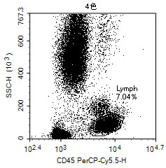
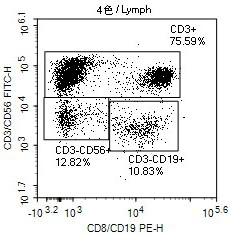
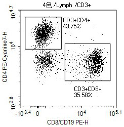
[0064] Using the kit in Example 1, a certain company's lymphocyte subset detection kit was used as a control kit to detect 20 clinical samples at the same time. In this example, two flow cytometers were used, namely NovoCyteD2060R and DxFLEX. The operation steps of each test are as follows: take 20 μL of monoclonal antibody mixture and 100 μL of peripheral blood and incubate at room temperature in the dark for 15 minutes, add 500 μL of red blood cell lysate and incubate at room temperature in the dark for 10 minutes, add 500 μL of PBS solution and incubate for 5 minutes at room temperature in the dark, that is, It can be checked on the machine. Repeat the above steps to obtain 80 sets of detection data, as shown in Table 1. Perform linear regression analysis on the test results, and compare the consistency of the test results of the two kits under different equipment. The results are shown in Table 2.

[0065] The data that table 1 embodiment 3 detects obtains

[0066]

...


PUM


Abstract

The invention discloses a kit for quantitatively detecting lymphocyte subsets and a detection method thereof. The kit provided by the invention comprises a mixture of six monoclonal antibodies, namelyCD3 FITC, CD56 FITC, CD45 PerCP-Cy5.5, CD8 PE, CD19 PE and CD4 PE-Cyanine7, and a red blood cell lysis buffer. The detection method comprises the following steps: taking a monoclonal antibody mixturein the kit and 100 [mu]L of peripheral blood, incubating at room temperature in a dark place, adding the red blood cell lysis buffer, incubating at room temperature in a dark place, adding a PBS solution, incubating at room temperature in a dark place to obtain a detection sample, detecting the detection sample by an up-flow cytometer, and determining the percentage of each lymphocyte subset. Thekit provided by the invention can be used for quantitatively detecting lymphocyte subsets, can reduce the requirements on the model of a convective cytometer, and is simple to operate and wide in application range.

Description

technical field [0001] The invention relates to the technical field of immunological detection, in particular to a kit for quantitatively detecting lymphocyte subsets and a detection method thereof. Background technique [0002] The immune status of the body is an important indicator to measure whether the body is sick. At present, flow cytometry is mostly used to monitor the immune status of the body. The most important indicator is to detect the levels of T, B, and NK lymphocytes. The determination of lymphoid subgroups is helpful to understand the immune status of the body and the monitoring of some diseases. Various monoclonal antibodies can be combined with antigens on the surface of lymphocytes, combined with multi-color fluorescent dyes, and various Lymphoid subgroups with different functions were distinguished, and then the relative ratio of each subgroup was obtained. CD45 molecules are expressed in all leukocytes, so the total lymphocytes are circled by CD45+, and...

Claims


Application Information

Patent Timeline
IPC IPC(8): G01N33/569G01N33/577G01N15/14
CPCG01N15/14G01N33/56966G01N33/577G01N2015/1006
Inventor 李国平李文娟
Owner UB BIOTECHNOLOGY ZHEJIANG CO LTD
Why Eureka
Try Eureka