Kit for detecting nuclear factor-kappa B and application of kit

A nuclear factor and kit technology, applied in the field of nuclear factor-κB detection kits, can solve the problems of low sensitivity, complicated design, and long time consumption, and achieve high sensitivity, low detection limit, and overcome interference

Active Publication Date: 2020-12-22
JIANGSU INST OF NUCLEAR MEDICINE
View PDF4 Cites 3 Cited by
  • Summary
  • Abstract
  • Description
  • Claims
  • Application Information

AI Technical Summary

Problems solved by technology

[0006] Therefore, some current signal amplification strategies based on molecular beacons to detect proteins have disadvantages such as low sensitivity, time-consuming, and overly complicated design.

Method used


Image

  • Kit for detecting nuclear factor-kappa B and application of kit
  • Kit for detecting nuclear factor-kappa B and application of kit
  • Kit for detecting nuclear factor-kappa B and application of kit

Examples


Embodiment 1

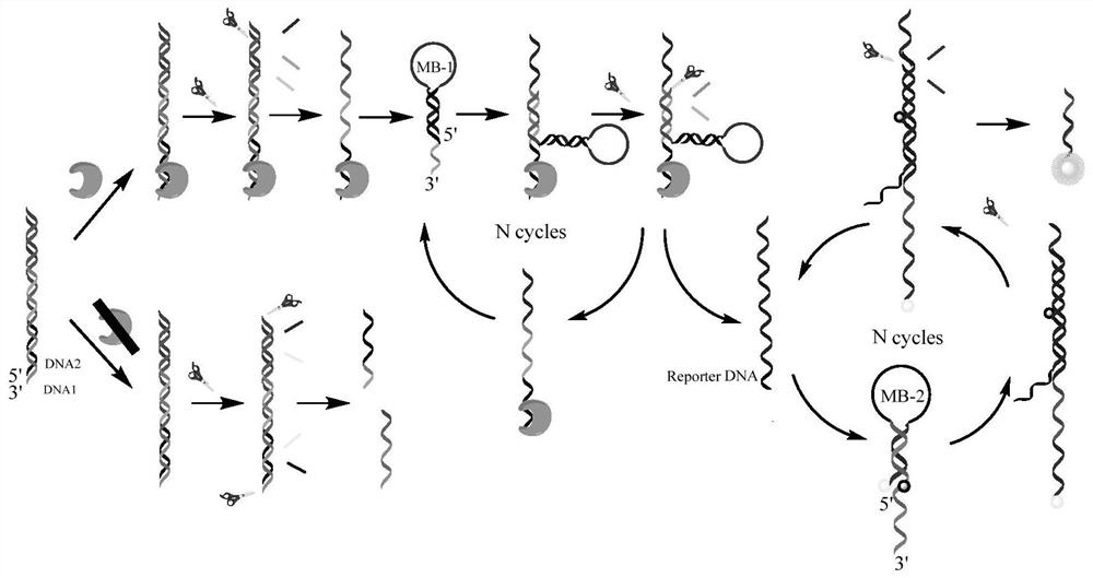
[0042] Mix DNA-1 (SEQ ID No.1) dissolved in DNA reaction buffer and DNA-2 (SEQ ID No.2) dissolved in DNA reaction buffer to obtain a double-stranded probe stock solution with a concentration of 5 μM . Prepared by dissolving molecular beacons MB-1 (SEQ ID No.3) and MB-2 (the 5' end of SEQ ID No.4 is marked with Cy3, and the 20th position is marked with BHQ-2) in DNA reaction buffer Both MB-1 and MB-2 concentrations were 5 μM stock solution. All solutions were heated at 95°C for 5 minutes, then slowly cooled to room temperature and allowed to stand for at least 4 hours.

[0043] When detecting NF-κB p65, add NF-κB p65 to 20uL protein binding buffer system, then add 40μL double-stranded DNA probe solution, and incubate at room temperature for 45min. Then add 20 μL Exo III at a concentration of 2.5U / μL, 40 μL MB-1 and 80 μL MB-2, the total volume of the system is 200 μL, the concentration of double-stranded DNA probe in this system is 1 μM, and the concentration of MB-1 is 1 μM ...

Embodiment 2

[0052] Embodiment 2 Sensitivity experiment

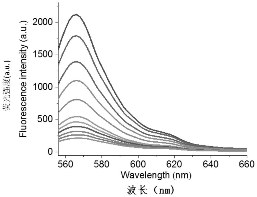
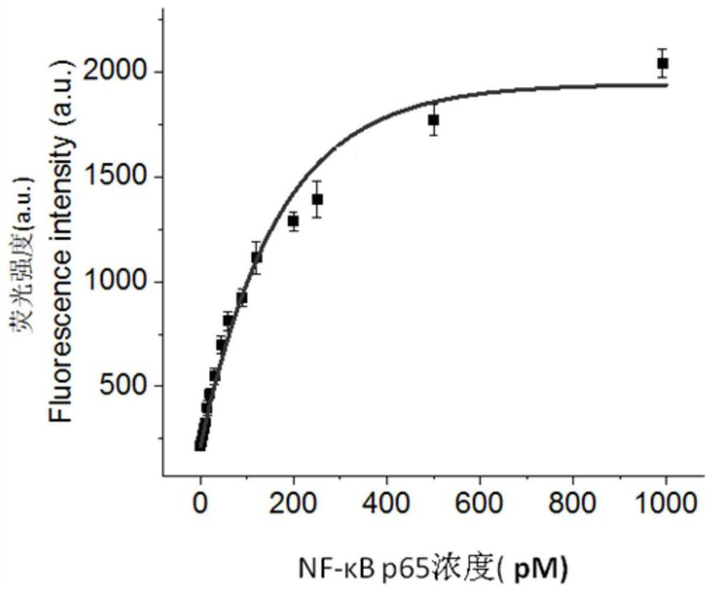
[0053] According to the method in Example 1, when detecting NF-κB p65, add NF-κB p65 to 20 uL protein binding buffer system, then add 40 μL double-stranded DNA probe solution, and incubate at room temperature for 45 min. Then add 20 μL 2.5U / μL ExoIII, 40 μL MB-1 and 80 μL MB-2, the concentration of double-stranded DNA probe in this system is 1 μM, the concentration of MB-1 is 1 μM, the concentration of MB-2 is 2 μM, and Incubate at 37°C for 1 h.

[0054] Detect the fluorescence spectrum of NF-κB p65 at different concentrations (0, 5, 10, 15, 20, 30, 60, 120, 250, 500 and 1000pM) in the range of 556-660nm with a multi-functional microplate reader, the results are shown in figure 2 . From figure 2 It can be seen that the fluorescence signal increases with the increase of NF-κB p65 concentration ( figure 2 The middle curve corresponds to the detection results of NF-κB p65 concentrations of 0, 5, 10, 15, 20, 30, 60, 120, 250, 500...

Embodiment 3

[0055] Embodiment 3 Feasibility study

[0056] Divided into 4 groups:

[0057] Group a: According to the method of Example 1, add 40 μL double-stranded DNA probe solution to 20 μL protein binding buffer system, and incubate at room temperature for 45 minutes; then add 40 μL MB-1 and 80 μL MB-2, and use DNA reaction buffer to complete the system Supplement to 200 μL, the concentration of double-stranded DNA probe in this system is 1 μM, the concentration of MB-1 is 1 μM, and the concentration of MB-2 is 2 μM, incubate at 37°C for 1 hour, and detect the concentration of the system in the range of 556-660nm Fluorescence spectrum; see results in Figure 4 curve a;

[0058] Group b: According to the method of Example 1, NF-κB p65 was added to 20 uL of protein binding buffer system, and then 40 μL of double-stranded DNA probe solution was added, and incubated at room temperature for 45 min. Add 40 μL MB-1 and 80 μL MB-2, and supplement the system to 200 μL with DNA reaction buffe...


PUM


Abstract

The invention provides a probe capable of detecting a nuclear factor-kappa B with high sensitivity, good specificity and short consumed time. The probe is a DNA double-stranded probe; the DNA double-stranded probe is formed by reverse complementation of DNA1 and DNA2; and the nucleotide sequence of the DNA1 is as shown in SEQ ID No. 1, and the nucleotide sequence of the DNA2 is as shown in SEQ IDNo. 2. The invention develops a simple, convenient and sensitive nuclear factor kappa B detection method. According to the method, DNA binding protein, exonuclease III (ExoIII) and an isothermal indexamplification technology are combined, and dual amplification of signals is successfully realized by adopting a molecular beacon dependent amplification fluorescence analysis technology. Compared with other methods, the method has higher specificity and lower detection limit, and can be directly used for detecting the nuclear factor kappa B in a cancer cell nuclear extracting solution.

Description

technical field [0001] The invention relates to the field of biotechnology, in particular to a kit for detecting nuclear factor-κB and its application. Background technique [0002] At present, severe acute respiratory syndrome coronavirus (SARS-CoV-2) has swept the world, causing serious harm and impact on human health. The autopsy results showed that the pathological features of patients with new coronary pneumonia (COVID-19) suggested the occurrence of acute respiratory distress syndrome (ARDS), and the cause of ARDS was cytokine storm. When the virus invades cells, the release of nucleic acid RNA activates Toll-like receptor 7 (TLR7) and recruits multiple proteins to form a complex, thereby promoting transcription factors (TFs) such as interferon regulatory factor 7 (IRF7) and nuclear factor-κB ( NF-κB) enters the nucleus and then activates the expression of pro-inflammatory cytokines, causing the immune system to become overactive, causing a cytokine storm, which is of...

Claims


Application Information

Patent Timeline
Patent Type & Authority Applications(China)
IPC IPC(8): G01N33/68G01N33/53G01N21/64C12Q1/6876C12Q1/6804C12N15/11
CPCG01N33/6872G01N33/53G01N21/6486C12Q1/6876C12Q1/6804C12Q2521/319C12Q2522/101C12Q2563/107Y02A50/30
Inventor 徐栋彭莹张凯徐希杰唐婕邹美芬钦晓峰
Owner JIANGSU INST OF NUCLEAR MEDICINE
Why Eureka
Try Eureka