Brain tumor multi-target auxiliary diagnosis and prospective treatment evolution visualization method and system

An auxiliary diagnosis and brain tumor technology, applied in the field of medical image processing, can solve the problems of lack of multi-target treatment evaluation model, poor classification results, insufficient data, etc.

Pending Publication Date: 2021-02-12
AFFILIATED HUSN HOSPITAL OF FUDAN UNIV
View PDF0 Cites 5 Cited by
  • Summary
  • Abstract
  • Description
  • Claims
  • Application Information

AI Technical Summary

Problems solved by technology

[0004] Although these models play a certain role in promoting the assistance of brain tumors, these models have certain deficiencies: (a) problems such as insufficient data lead to fewer researches on auxiliary diagnosis based on multi-disease classification, and the classification results are poor; ( b) There is a lack of multi

Method used


Image

  • Brain tumor multi-target auxiliary diagnosis and prospective treatment evolution visualization method and system
  • Brain tumor multi-target auxiliary diagnosis and prospective treatment evolution visualization method and system
  • Brain tumor multi-target auxiliary diagnosis and prospective treatment evolution visualization method and system

Examples


Embodiment 1

[0092] In order to construct a prospective visualization model of brain tumor multi-target auxiliary diagnosis and brain tumor treatment growth evolution that can meet the needs of clinical applications, the present invention proposes a brain tumor multi-target auxiliary diagnosis and prospective treatment evolution visualization method. To overcome the shortcomings of the existing brain tumor auxiliary diagnosis and treatment technology.

[0093] According to the present invention, a brain tumor multi-target auxiliary diagnosis and prospective treatment evolution visualization method includes:

[0094] Step M1: Acquire the multi-target multi-modal MRI data of brain tumors before and after treatment, and preprocess the multi-target multi-modal MRI data of brain tumors before and after treatment to obtain a unified standard before and after treatment. Paired Brain Tumor Multitarget Multimodal MRI Data I original and I later ;

[0095] Step M2: Multi-target and multi-modal MR...

Embodiment 2

[0133] Embodiment 2 is a modification of embodiment 1

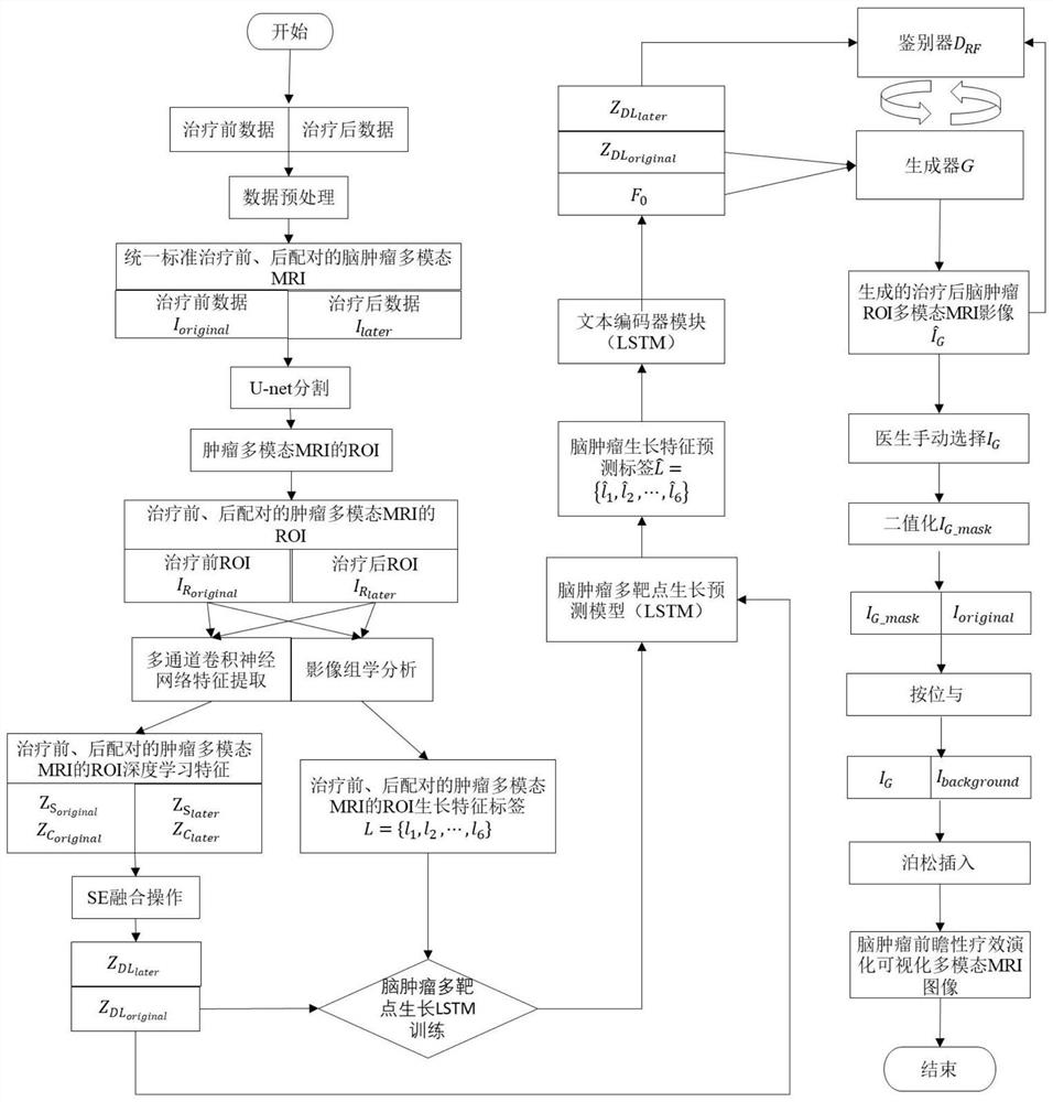
[0134] figure 1 As shown in the present invention, a brain tumor multi-target auxiliary diagnosis and prospective treatment evolution visualization method includes the following steps:

[0135]Step (1): Brain tumor multi-target and multi-modal MRI data (including: T1, T1C, T2, Flair, PWI and ADC) preprocessing; according to the latest WHO recommendations, clinical guidelines and pathological data, the doctor calibrates the brain tumor multiple Multiple molecular gene categories of modal MRI data: IDH-mutant / wildtype, 1p / 19qCo-Deletion, EGFR, PTEN), TERT, p53TP53, ATRX, and ALK, etc.; The processing method obtains the multi-target and multi-modal MRI data of brain tumors with uniform resolution and approximately the same gray distribution before and after treatment, respectively recorded as I original and I later , and the size of the .nii.gz data for each modality is 256×256×16.

[0136] Step (2): brain tumor ROI mult...


PUM


Abstract

The invention provides a brain tumor multi-target auxiliary diagnosis and prospective treatment evolution visualization method and system. The brain tumor multi-target auxiliary diagnosis and prospective treatment evolution visualization method comprises the steps: acquiring and preprocessing paired brain tumor multi-target multi-mode MRI data before and after treatment; carrying out tumor regionsegmentation on the preprocessed paired brain tumor multi-target multi-mode MRI data before and after treatment through a 3DU-net convolutional neural network, and obtaining a growth characteristic label L = {l1, l2, l3,..., ln} through an imaging omics method; and performing feature extraction on the sum through a multi-channel convolutional neural network and then performing SE fusion operationto obtain deep learning features and input the deep learning features into a prediction model to obtain a brain tumor multi-target growth prediction label, inputting the brain tumor multi-target growth prediction label into a trained prospective treatment visualization model to obtain a final brain tumor region-of-interest growth evolution image, and inserting the brain tumor region-of-interest growth evolution image into a non-brain tumor region Ibackground to complete a brain tumor prospective treatment visualization task. The method and system in the invention has higher clinical practicability.

Description

technical field [0001] The present invention relates to the technical field of medical image processing, in particular to a method and system for visualization of multi-target auxiliary diagnosis and prospective treatment evolution of brain tumors. Background technique [0002] Brain tumors are common tumors in the human body. Central neurologists use information such as multimodal magnetic resonance imaging (MRI) of the patient's brain to diagnose the patient's illness. At present, the gold standard for accurate diagnosis of brain tumors is still histopathological examination and genetic testing, but tumor biopsy is an invasive operation, the operation risk is high, and it cannot accurately reflect the internal heterogeneity of tumor tissue; in the era of medical digitalization , Computer Assisted Diagnosis (CAD) technology can combine multimodal MRI images of the brain (including: T1-weighted imaging (T1), T1-weighted enhanced imaging (T1C), T2-weighted imaging (T2), water...

Claims


Application Information

Patent Timeline
IPC IPC(8): G16H50/30G06T7/00G06T7/11G06T7/45G06T7/62G06K9/32G06K9/62G06N3/04G06N3/08
CPCG16H50/30G06T7/0012G06T7/11G06T7/45G06T7/62G06N3/049G06N3/08G06T2207/10088G06T2207/20081G06T2207/20084G06T2207/20221G06T2207/30016G06T2207/30096G06V10/25G06N3/045G06F18/253
Inventor 于泽宽耿道颖刘晓曹鑫李郁欣刘军张军尹波刘杰吴昊耿岩胡斌张海燕杜鹏陆逸平
Owner AFFILIATED HUSN HOSPITAL OF FUDAN UNIV
Why Eureka
Try Eureka