3D printed femoral head necrosis navigation template and construction method thereof and use of navigation template and construction method

A femoral head necrosis and navigation template technology, applied in surgical navigation systems, computer-aided planning/modeling, medical science, etc., can solve problems such as high operator experience requirements, large patient trauma, and large radiation of medical staff, and reduce Radiation damage, easy-to-master effects

Pending Publication Date: 2021-02-23
中国人民解放军联勤保障部队第九二〇医院
View PDF0 Cites 5 Cited by
  • Summary
  • Abstract
  • Description
  • Claims
  • Application Information

AI Technical Summary

Problems solved by technology

The previous method is to use body surface positioning, X-ray multi-directional fluoroscopy, insert a guide pin, and then fluoroscopy again to determine the angle, depth, and direction, and then expand the tunnel to remove necrotic bone. Preoperative planning, accurate positioning, at the same time, during the operation, the medical staff and patients receive a lot of radiation, and repeated operations to determine the angle and depth will cause greater trauma to the patient, and at the same time require a higher level of experience for the operator , it is not convenient for technology mastering and technology promotion
[0004] To sum up, the existing technology currently lacks an individual-customized navigation template for femoral head necrosis for the treatment of femoral head necrosis, and lacks a corresponding navigation template construction method

Method used


Image

  • 3D printed femoral head necrosis navigation template and construction method thereof and use of navigation template and construction method
  • 3D printed femoral head necrosis navigation template and construction method thereof and use of navigation template and construction method
  • 3D printed femoral head necrosis navigation template and construction method thereof and use of navigation template and construction method

Examples


Embodiment 1

[0043] Such as figure 2 shown

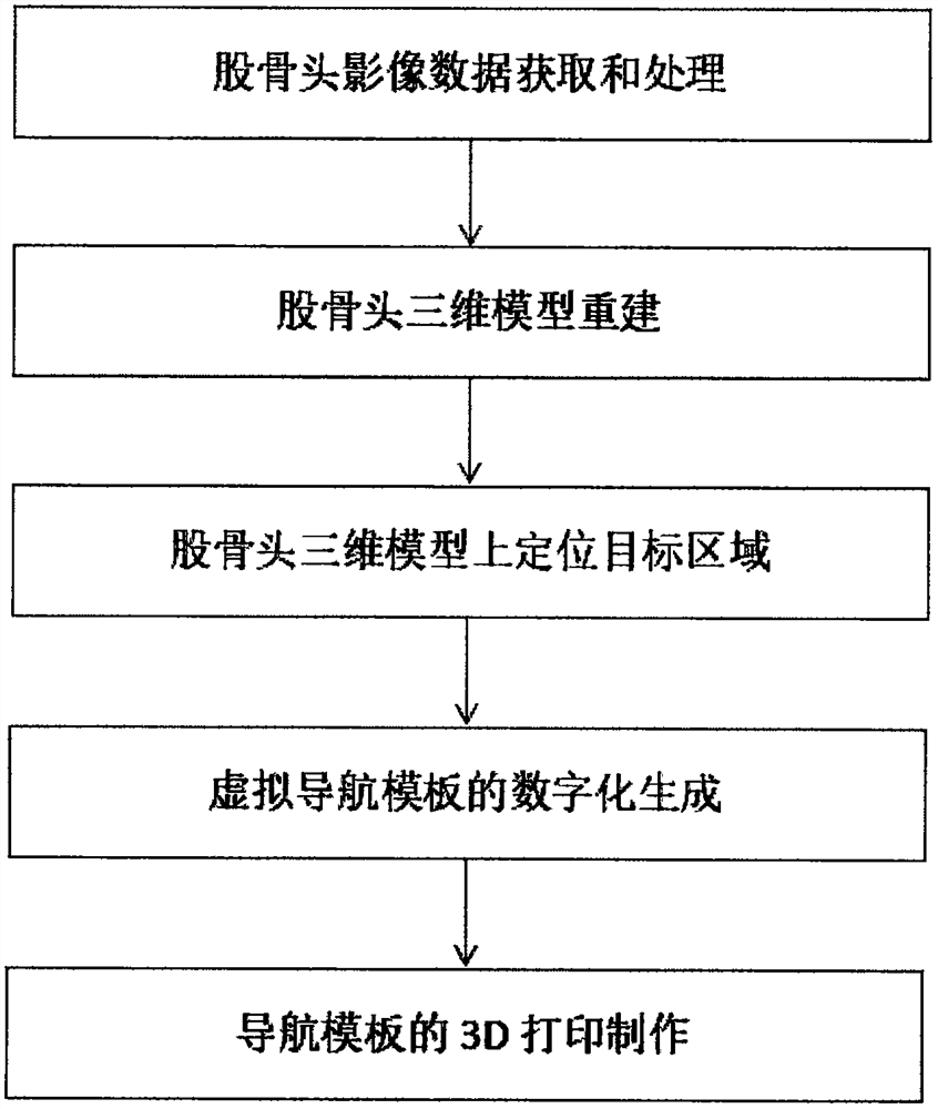
[0044] A construction method of a 3D printed femoral head necrosis navigation template, comprising the following steps:

[0045] Step 1. Femoral head image data acquisition and processing

[0046] A 64-slice spiral CT was used for thin-slice scanning of the hip joint, and the original CT image data was imported into Mimics 15.0 software in Dicom format;

[0047] Step 2. Reconstruction of the 3D model of the femoral head

[0048] In Mimics 15.0 software, use threshold segmentation (Thresholding) and region growing (Regiongrowing) to obtain the mask (Mask), and use the editing mask tool (Editing masks) to carefully separate the acetabulum and femoral head layer by layer, and erase the hip joint , keep the femoral head, use region growth to extract the femur, save it as a new femoral mask, use the Boolean operation function to complete the separation of the acetabular side, and calculate the 3D model ( Calculate 3D) to complete the three-dimen...

Embodiment 3

[0061] Such as Figure 3-6 shown

[0062] Animal experiments of the construction of 3D printed femoral head necrosis navigation template

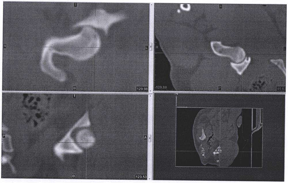
[0063] data collection

[0064] Random number table method was used to select Beagle dogs, using pentobarbital sodium salt (Merck Chemical Technology (Shanghai) Co., Ltd., China) to prepare 3% pentobarbital sodium solution, 1ml / kg intravenous anesthesia, after the anesthesia was stable The prone position was fixed on a 64-slice spiral CT (GE Medieal Systems, USA) platform, and the bilateral hip joints of Beagle dogs were scanned with thin layers. Scanning data setting: voltage 120KV, current 150MA, matrix 512*512, layer thickness 0.625mm. Raw CT image data were imported into Mimics 15.0 software (Materialise Corporation, Belgium) in Dicom format. ( image 3 )

[0065] Three-dimensional model reconstruction of canine femoral head

[0066] Using threshold segmentation (Thresholding) in Mimics 15.0 software, segment along the gray value...

Embodiment 4

[0080] Application of 3D printed femoral head necrosis navigation template and construction method in femoral head necrosis localization and navigation.

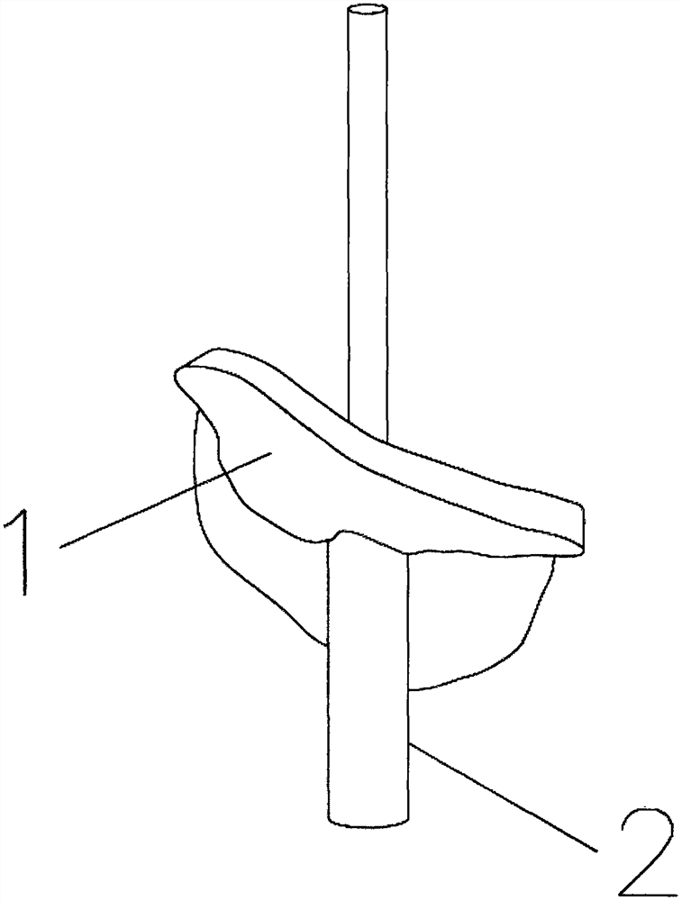
[0081] According to the specific description of the above-mentioned embodiment, the navigation template for precise positioning is customized through preoperative planning, that is, through the acquisition and processing of femoral head image data, the reconstruction of the three-dimensional model of the femoral head, the positioning of the target area on the three-dimensional model of the femoral head, and the digital generation of the virtual navigation template , 3D printing of the navigation template to obtain a navigation base template that can accurately match the greater trochanter of the femoral head, and a navigation tube with the best needle insertion angle and orientation, which can be customized and planned surgical operations, and can be intuitively applied to surgical operations Navigation can effectively reduce...


PUM


Abstract

The invention discloses a 3D printed femoral head necrosis navigation template and a construction method thereof and use of the navigation template and construction method. The construction method comprises the following steps: acquiring and processing femoral head image data, reconstructing a femoral head three-dimensional model, positioning a target area on the femoral head three-dimensional model, digitally generating a virtual navigation template, and performing 3D printing on the navigation template to obtain a navigation base template which can be accurately matched with a femoral head greater trochanter area and a navigation tube with an optimal needle insertion angle and direction. The femoral head necrosis navigation template obtained on the basis of the three-dimensional reconstruction and 3D printing can be visually applied to operation navigation of an operation, effectively reduces radiation injuries and wounds to a patient, and is convenient for an operator to master, operate and perform technology promotion.

Description

technical field [0001] The present invention relates to the technical field of a femoral head necrosis positioning and navigation device, in particular to a 3D printed femoral head necrosis navigation template and its construction method and application. Background technique [0002] Avascular necrosis of the femoral head is a common clinical disease. There are many treatment methods for avascular necrosis of the femoral head. Among them, drilling and decompression of the femoral head was first used by the Germans to treat avascular necrosis of the femoral head. The early effect is remarkable. In theory , Reduce intraosseous pressure, rebuild blood circulation, restore blood flow to the femoral head, make dead bone tissue have the opportunity to contact living tissue, and promote the repair process. Core decompression in the treatment of early avascular necrosis of the femoral head has definite curative effect, which has been recognized by most clinical orthopedic surgeons. ...

Claims


Application Information

Patent Timeline
Patent Type & Authority Applications(China)
IPC IPC(8): A61B34/10A61B34/20A61B17/56B33Y10/00B33Y30/00B33Y50/00B33Y70/00B33Y80/00
CPCA61B17/56A61B2017/00526A61B2017/564A61B34/10A61B34/20A61B2034/105A61B2034/108A61B2034/2068B33Y10/00B33Y30/00B33Y50/00B33Y70/00B33Y80/00
Inventor 冯凡哲徐永清何晓清陆声罗浩天
Owner 中国人民解放军联勤保障部队第九二〇医院
Why Eureka
Try Eureka