An automatic analysis method for deformed long bone shape

An automatic analysis and deformity technology, which is applied in the field of medical image processing, can solve problems such as inaccurate positioning point marking, inaccurate measurement angle readings, and line deviation errors, etc., to reduce the burden on doctors, good repeatability, and time-consuming Excellent effect

An automatic analysis and deformity technology, which is applied in the field of medical image processing, can solve problems such as inaccurate positioning point marking, inaccurate measurement angle readings, and line deviation errors, etc., to reduce the burden on doctors, good repeatability, and time-consuming Excellent effect

CN112435255BActive Publication Date: 2022-04-29HEBEI UNIV OF TECH

Image

  • An automatic analysis method for deformed long bone shape
  • An automatic analysis method for deformed long bone shape
  • An automatic analysis method for deformed long bone shape

Examples


Embodiment 1

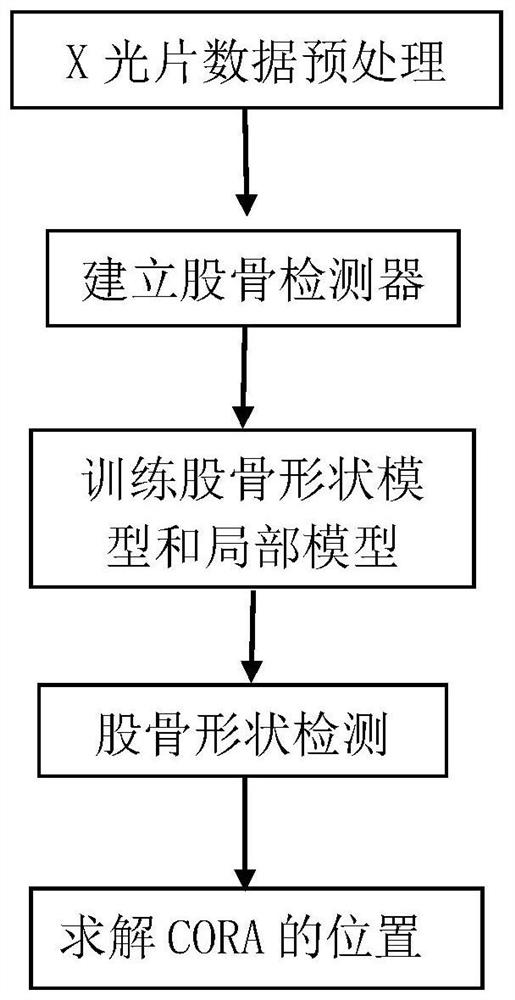
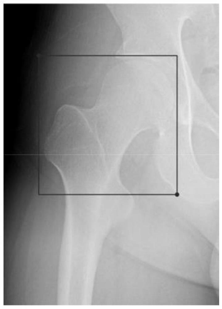
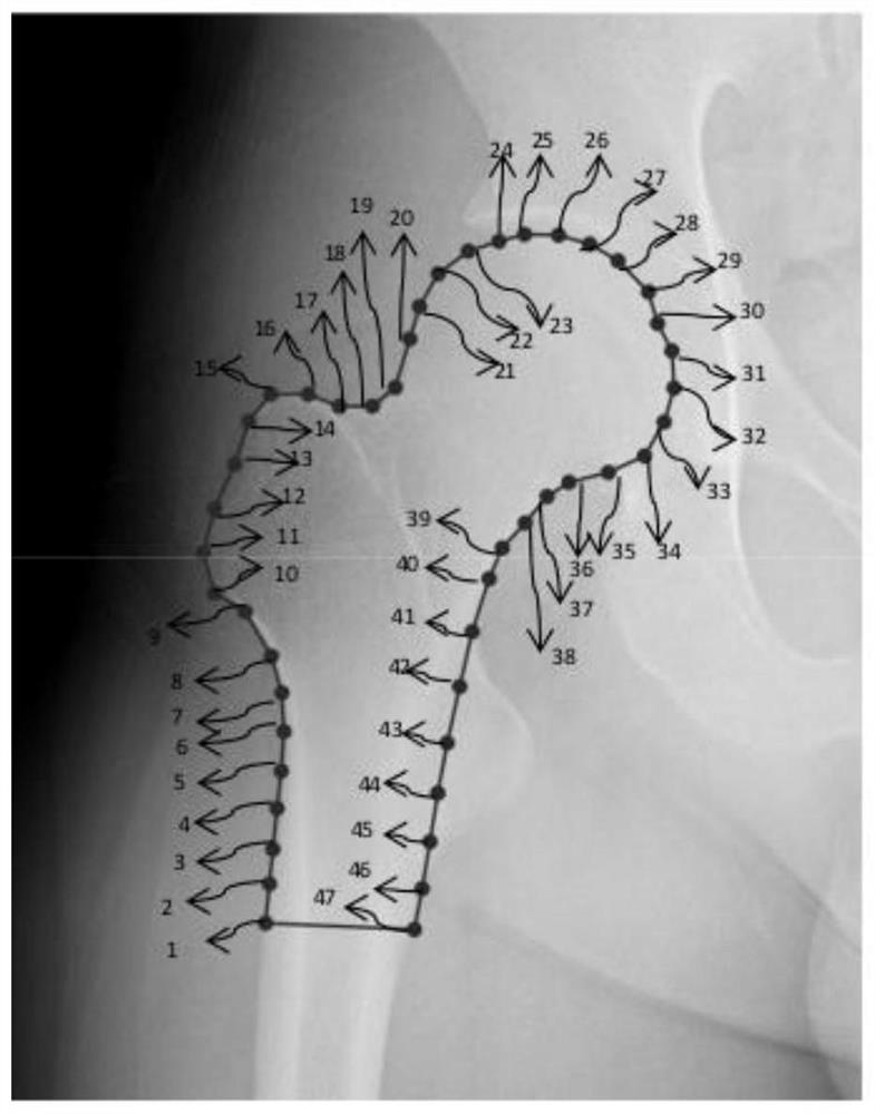
[0144] Using the analysis method of the present invention to analyze the shape of deformed long bones, a total of 120 X-ray films of deformed long bones are used, 100 of which are training sets, and the remaining 20 are test sets. Under the guidance of a doctor, the position of the femur and the feature points of the femur are marked manually. First, mark the X-rays in the training set, use a rectangular frame to mark the proximal femur and the distal end of the bone, use 47 points to mark the contour of the proximal femur, and use 49 points to mark the contour of the distal femur, as shown in Figure 2-5 As shown, the points with the same serial number were basically located at the same position of the femur during the marking process. According to the operations of the first step to the fourth step of the analysis method of the present invention, the optimal position of the contour feature point of the proximal end of the femur and the optimal position of the contour feature...


PUM


Abstract

The invention discloses an automatic analysis method for deformed long bone shape. In the analysis method, a femur detector, a shape model and a local model are trained according to a femur X-ray film set. The position and size of the femur in the X-ray film are detected by the trained detector. The position and size are used as the initial values ​​of the trained shape model to fit the femur contour. Finally, according to the position of each feature point after fitting, the anatomical axis of each bone segment of the femur and the joint line are drawn. When this method is used for measurement, it is almost consistent with manual measurement results, and the time-consuming is better than traditional measurement methods. Applying the analysis method of the present invention for automatic deformity analysis can make the most effective use of image data, avoid image deformation, automatic line drawing, measurement and other functions can eliminate manual errors such as line drawing errors, protractor errors, reading errors, etc., and the repeatability is good. The analysis method of the invention can meet the accuracy requirement of orthopedics, can significantly reduce the burden on doctors, and improve work efficiency.

Description

technical field [0001] The invention relates to the technical field of medical image processing, in particular to an automatic analysis method for deformed long bone shape. Background technique [0002] Taylor Spatial Frame (TSF) is one of the latest advances in the field of deformity correction. It utilizes the principle of Stewart platform and is a representative of hexapod robot external fixator. Correction of the deformity requires an osteotomy, which requires the determination of the center of rotation of angle (CORA)—the angle at which the proximal axis and the distal axis intersect. Analyzing the deformity on the X-ray film and drawing the anatomical axis of each bone segment is the primary task of deformity correction with Taylor space external fixator. The current method of deformity analysis is to place the X-ray film on the light box, use a crayon or a marker pencil to mark directly on the X-ray film, and then use a protractor to measure the angle between the axe...

Claims


Application Information

Patent Timeline
29 Apr 2022
Publication
CN112435255B
IPC
G06T7/00; G06T7/11; G06V10/774; G06V10/764; G06K9/62
CPC
G06T7/0012; G06T7/11; G06T2207/10116; G06T2207/20021; G06T2207/20081; G06T2207/30008; G06V2201/07; G06F18/2411
Inventors
孙昊; 刘德龙