
Method and device for extracting pulmonary embolism image, storage medium and electronic equipment

A pulmonary embolism and image technology, applied in the field of image processing, can solve problems such as huge workload and difficult collection of CTPA image samples

Pending Publication Date: 2021-03-09
沈阳东软智能医疗科技研究院有限公司
View PDF14 Cites 2 Cited by
  • Summary
  • Abstract
  • Description
  • Claims
  • Application Information

AI Technical Summary

Problems solved by technology

However, this method requires a large amount of sample data of marked embolized areas, and the workload of manually marking embolized areas is huge
Moreover, it is difficult to collect a variety of CTPA image samples

Method used


Image

  • Method and device for extracting pulmonary embolism image, storage medium and electronic equipment
  • Method and device for extracting pulmonary embolism image, storage medium and electronic equipment
  • Method and device for extracting pulmonary embolism image, storage medium and electronic equipment

Examples


Embodiment Construction

[0077] Specific embodiments of the present disclosure will be described in detail below in conjunction with the accompanying drawings. It should be understood that the specific embodiments described here are only used to illustrate and explain the present disclosure, and are not intended to limit the present disclosure.

[0078] Reference will now be made in detail to the exemplary embodiments, examples of which are illustrated in the accompanying drawings. When the following description refers to the accompanying drawings, the same numerals in different drawings refer to the same or similar elements unless otherwise indicated. The implementations described in the following exemplary examples do not represent all implementations consistent with the present disclosure. Rather, they are merely examples of apparatuses and methods consistent with aspects of the present disclosure as recited in the appended claims.

[0079] In order to make it easier for those of ordinary skill i...


PUM


Abstract

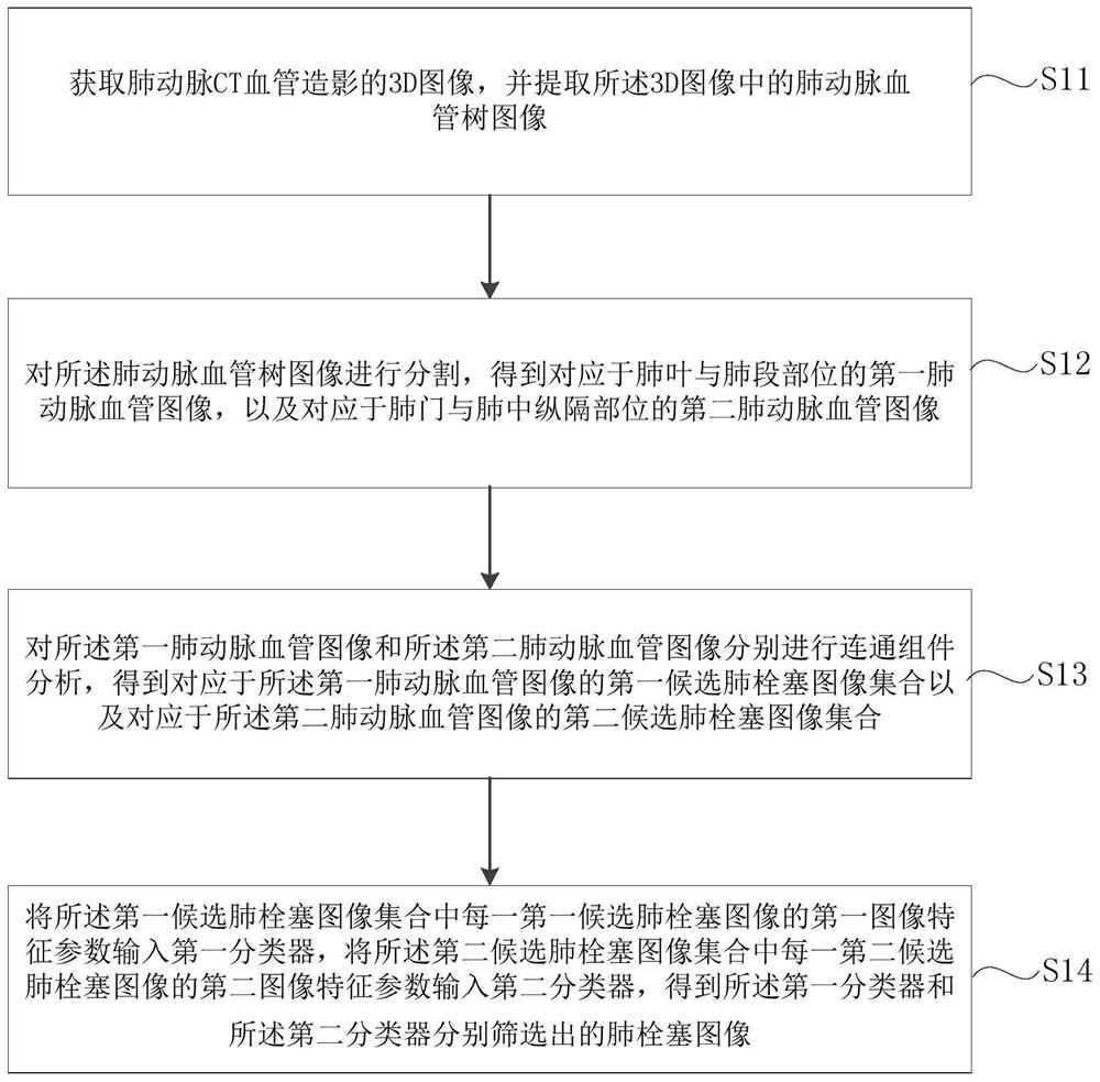
The invention relates to a method and device for extracting a pulmonary embolism image, a storage medium and electronic equipment, and aims to accurately extract the pulmonary embolism image. The method comprises the steps of obtaining a 3D image of pulmonary artery CT angiography, and extracting a pulmonary artery blood vessel tree image in the 3D image; segmenting the pulmonary artery blood vessel tree image to obtain a first pulmonary artery blood vessel image corresponding to the pulmonary lobe and lung segment part and a second pulmonary artery blood vessel image corresponding to the pulmonary portal and lung medial septum part; performing connected component analysis on the first pulmonary artery blood vessel image and the second pulmonary artery blood vessel image to obtain a firstcandidate pulmonary embolism image set and a second candidate pulmonary embolism image set; and inputting the first image feature parameter of each first candidate pulmonary embolism image into a first classifier, and inputting the second image feature parameter of each second candidate pulmonary embolism image into a second classifier to obtain pulmonary embolism images screened by the first classifier and the second classifier respectively.

Description

technical field [0001] The present disclosure relates to the technical field of image processing, and in particular, to a method, device, storage medium and electronic equipment for extracting images of pulmonary embolism. Background technique [0002] Pulmonary embolism (PE for short) refers to the clinical pathophysiological syndrome of pulmonary circulation disorder caused by various emboli in the systemic circulation blocking the pulmonary artery and its branches. Patients with pulmonary embolism will suddenly experience unexplained collapse, pale complexion, cold sweat, dyspnea, chest pain, cough and other symptoms, as well as symptoms of cerebral hypoxia such as extreme anxiety, fatigue, nausea, convulsions, and coma. Pulmonary embolism is a common disease with high morbidity and mortality, and its incidence is second only to acute myocardial infarction and aortic dissection. However, due to the "atypical" nature of symptoms and signs of pulmonary embolism, it is easy...

Claims


Application Information

Patent Timeline
Patent Type & Authority Applications(China)
IPC IPC(8): G06T7/00G06T7/11G06T7/136G06K9/34G06K9/62G06N3/04G06N3/08
CPCG06T7/0012G06T7/11G06T7/136G06N3/08G06T2207/10081G06T2207/20081G06T2207/20084G06T2207/30061G06V10/267G06N3/045G06F18/241
Inventor 平安何光宇王希
Owner 沈阳东软智能医疗科技研究院有限公司
Learn More