
Method for detecting directional distribution of human mesenchymal stem cells in animal body

A technology of mesenchymal stem cells and detection methods, which is applied in the field of detection of the directional distribution of implanted human mesenchymal stem cells in traced animals, and can solve problems such as the observation of lagging overall function improvement in stem cell labeling research

Pending Publication Date: 2021-03-16
YUNNAN SHUNXI REGENERATION MEDICAL ENG CO LTD
View PDF0 Cites 1 Cited by
  • Summary
  • Abstract
  • Description
  • Claims
  • Application Information

AI Technical Summary

Problems solved by technology

However, the tracer studies of stem cell markers to evaluate cell engraftment, distribution, survival, migration, differentiation and function obviously lag behind the observation of overall functional improvement, which has become a bottleneck factor and a new research hotspot in the investigation of stem cell therapy mechanism

Method used


Image

  • Method for detecting directional distribution of human mesenchymal stem cells in animal body
  • Method for detecting directional distribution of human mesenchymal stem cells in animal body
  • Method for detecting directional distribution of human mesenchymal stem cells in animal body

Examples


Embodiment 1

[0131] This embodiment provides a method for detecting the directional distribution of human-derived mesenchymal stem cells in animals, specifically the detection of the number of hPC-MSC human placental chorionic mesenchymal stem cells in peripheral blood samples of cynomolgus monkeys;

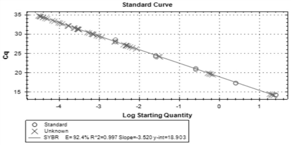
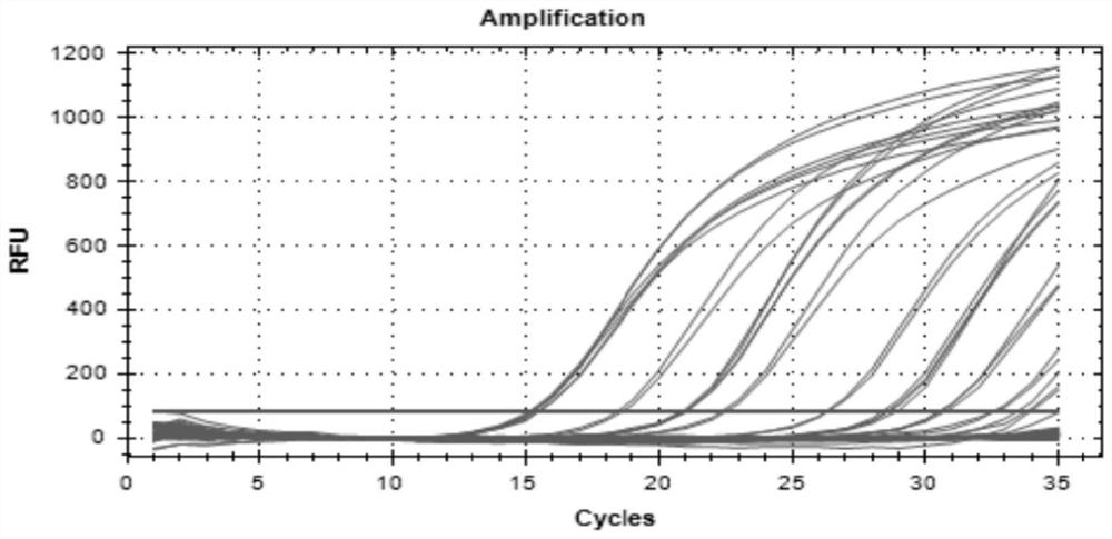
[0132] Methods: Fluorescent quantitative PCR was used to detect the DNA concentration of hPC-MSC human placental chorionic mesenchymal stem cell injection in the peripheral blood of cynomolgus monkeys collected in the experiment, and calculate the cell number;

[0133]The applicant collected 512 blood samples and 190 tissue samples from 2019.04.23 to 2019.08.14, a total of 702, and stored them in a refrigerator below -66°C. The DNA extraction was completed from 2019.08.14 to 2019.11.11. 2019.09.17-2019.11.13 completed sample qPCR detection;

[0134] Results: Under the experimental research dose, after the first administration of each dose group, the DNA concentration of hPC-MSC human placenta...

Embodiment 2

[0161] This embodiment provides a detection method for the directional distribution of human-derived mesenchymal stem cells in animals, specifically: detection of the number of hPC-MSC human placental chorionic mesenchymal stem cells in cynomolgus monkey tissue samples;

[0162] Methods: Fluorescent quantitative PCR was used to detect the DNA concentration of hPC-MSC human placental chorionic mesenchymal stem cell injection in the heart, liver, kidney, spleen, and lung tissues of cynomolgus monkeys collected in the experiment, and calculate the cell number

[0163] Results: Under the experimental study dose, no hPC-MSC human placental chorionic mesenchymal stem cell injection DNA was detected in each tissue sample at all time points.

[0164] Under the experimental research dose, DNA of hPC-MSC human placental chorionic mesenchymal stem cell injection was not detected in the tissue samples after intravenous infusion of 0.9% sodium chloride injection in cynomolgus monkeys (contr...


PUM

PropertyMeasurementUnit
concentrationaaaaaaaaaa

Abstract

The invention relates to a method for detecting directional distribution of human mesenchymal stem cells in an animal body, DNA concentration of hPCMSC human placenta chorionic mesenchymal stem cell injection on peripheral blood and organ tissue samples of a cynomolgus monkey collected in a test is detected by a fluorescence quantitative PCR method (Polymerase Chain Reaction) method, and the cellnumber is calculated. The method can be used for evaluating the safety after the human mesenchymal stem cells are implanted into the animal body. The method for tracing the directional distribution ofthe human mesenchymal stem cells implanted into the animal body is a cell distribution detection method which integrates the advantages of safety, sensitivity, specificity, non-invasiveness, persistence and the like into a whole in an animal experiment.

Description

technical field [0001] The invention belongs to the technical field of biomedicine, and in particular relates to a detection method for tracing the directional distribution of human-derived mesenchymal stem cells implanted in animals. Background technique [0002] Mesenchymal stem cells are a type of adult stem cells with strong self-renewal and replication capabilities and multi-directional differentiation potential; mesenchymal stem cells have low immunogenicity and antigen presentation capabilities, and have unique bidirectional regulatory capabilities in immune regulation; It has great clinical application value in graft-versus-host disease, autoimmune disease, cardiovascular system disease, neurodegenerative disease and so on. Currently, umbilical cord-placental-derived mesenchymal stem cells are increasingly becoming a hot source of mesenchymal stem cells. Neonatal umbilical cords are medical wastes, which are easy to collect, cause no harm to the donor, and do not in...

Claims


Application Information

Patent Timeline
Patent Type & Authority Applications(China)
IPC IPC(8): C12Q1/6851
CPCC12Q1/6851C12Q2531/113C12Q2545/113C12Q2537/165
Inventor 李春姜小锋陈曦闫姗姗
Owner YUNNAN SHUNXI REGENERATION MEDICAL ENG CO LTD
Learn More