
Multi-mode optical microscopic imaging device and microscopic imaging method

An optical microscope and imaging device technology, applied in medical imaging, medical science, analysis using fluorescence emission, etc., can solve the problem of failure to realize the simultaneous implementation of multiple optical imaging modes, limited integration of miniaturized microscopes, and inability to apply specific behavioral patterns etc.

Pending Publication Date: 2021-04-06
HUAZHONG PHOTOELECTRIC TECH INST (CHINA SHIPBUILDING IND CORP THE NO 717 INST)
View PDF4 Cites 2 Cited by
  • Summary
  • Abstract
  • Description
  • Claims
  • Application Information

AI Technical Summary

Problems solved by technology

However, limited by the degree of integration of miniaturized microscopes, current head-mounted microscopes can only achieve a single optical imaging mode, such as wide-field fluorescence imaging or two-photon fluorescence imaging, but fail to realize the simultaneous implementation of multiple optical imaging modes
In addition, the existence of cables still restricts the free movement process of experimental animals to a certain extent, and cannot be applied to some special behavior modes, such as predation, digging, etc.

Method used


Image

  • Multi-mode optical microscopic imaging device and microscopic imaging method
  • Multi-mode optical microscopic imaging device and microscopic imaging method

Examples


Embodiment Construction

[0021] The specific implementation manners of the present invention will be further described in detail below in conjunction with the accompanying drawings and embodiments. The following examples are used to illustrate the present invention, but are not intended to limit the scope of the present invention.

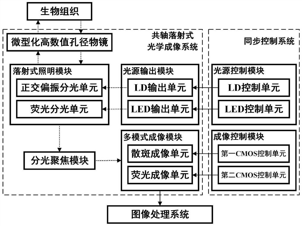
[0022] figure 1 A multi-mode optical micro-imaging device provided by the present invention, the micro-imaging device includes a coaxial epi-optical imaging system and an image processing system, wherein the co-axial epi-optical imaging system includes a light source output module, an epi-optical Type illumination module, miniaturized high numerical aperture objective lens, spectroscopic focusing module and multi-mode imaging module.

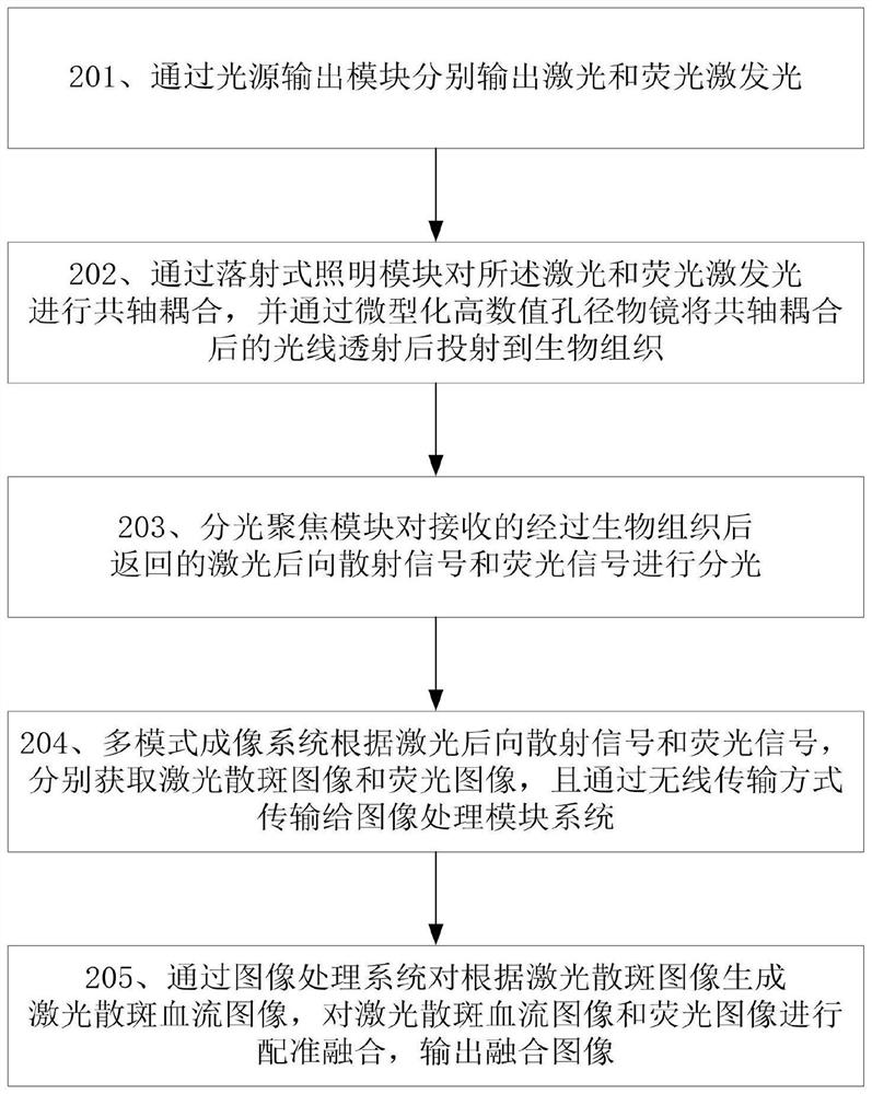
[0023]Wherein, the light source output module is used to output the laser light and the fluorescence excitation light respectively, and inject the epi-type illumination module; The light incident on the miniaturized high numerical aperture...


PUM


Abstract

The invention provides a multi-mode optical microscopic imaging device and a microscopic imaging method. The device comprises a coaxial incident optical imaging system and an image processing system, and the coaxial incident optical imaging system comprises a light source output module, an incident illumination module, a miniaturized high numerical aperture objective lens, a light splitting focusing module and a multi-mode imaging module. The method can be applied to an experimental animal in a waking free activity state, and high-resolution and stable blood flow change distribution and nerve activity distribution in the free behavior process of the experimental animal can be obtained at the same time; multiple imaging modes in the same view field area can be fused by adopting a registration fusion technology; based on the implantable miniaturized objective lens, superficial or deep tissue imaging of an experimental animal can be realized.

Description

technical field [0001] The invention relates to the field of biological optical imaging, and more specifically, to a multi-mode optical microscopic imaging device and a microscopic imaging method. Background technique [0002] Brain functional optical imaging technology, with its advantages of high temporal and spatial resolution and multi-parameter information acquisition, can study brain functional activities from different spatial and temporal scales, and plays a vital role in the field of neuroscience. Optical imaging of brain function in awake animals is one of the current development trends in optical imaging of brain function. For a long time, most of the traditional optical imaging methods of brain function have been applied to anesthetized animals, and only a few studies have been carried out based on awake animals. However, one of the main purposes of brain science research is to explain the mechanism of high-level neural activities such as perception and behavior...

Claims


Application Information

Patent Timeline
Patent Type & Authority Applications(China)
IPC IPC(8): A61B5/00
CPCA61B5/00A61B5/0042A61B5/0062A61B5/0071A61B5/0084A61B5/6868A61B2503/40A61B2503/42A61B2576/026
Inventor 陈铭徐寅李邦旭陈松操琼
Owner HUAZHONG PHOTOELECTRIC TECH INST (CHINA SHIPBUILDING IND CORP THE NO 717 INST)
Learn More