Cell preserving fluid, preparation method thereof and cell preserving method

A preservation method and preservation solution technology, applied in the preservation, application, animal husbandry, etc. of human or animal bodies, can solve the requirements that it is difficult to ensure the number and quality of effective cells, the epithelial cells cannot be precipitated, and mucus substances cannot be dissolved, etc. problems, to achieve the effect of preserving multiple cell types, protecting the working environment and staff health, and preventing cell aggregation

Active Publication Date: 2021-04-13
深路医学科技(武汉)有限公司
View PDF7 Cites 4 Cited by
  • Summary
  • Abstract
  • Description
  • Claims
  • Application Information

AI Technical Summary

Problems solved by technology

Cells are often wrapped in mucus, which affects the effect of cell production, and a large amount of mucus often leads to failure of production
At present, measures such as strengthening the shaking intensity, prolonging the shaking time, differential centrifugation, and freezing are often used clinically, but the effect is not obvious, and the mucus substance cannot be dissolved, and the epithelial cells in the mucus substance cannot be precipitated, so it is difficult to guarantee the detection. Requirements for effective cell quantity and quality are not conducive to clinical diagnosis

Method used


Image

  • Cell preserving fluid, preparation method thereof and cell preserving method
  • Cell preserving fluid, preparation method thereof and cell preserving method
  • Cell preserving fluid, preparation method thereof and cell preserving method

Examples


Embodiment 1-3

[0053] The formula of the cell preservation solution provided by Examples 1-3 is shown in Table 1 below:

[0054] Table 1

[0055] Example 1 Example 2 Example 3 Ethanol (95% pure) 40% (v / v) 45% (v / v) 50% (v / v) Tris 0.1% (w / v) 0.3% (w / v) 0.5% (w / v) EDTA·2Na 0.01% (w / v) 0.02% (w / v) 0.03% (w / v) bromelain 0.01% (w / v) 0.05% (w / v) 0.04% (w / v) Tris(2-carboxyethyl)phosphine hydrochloride 0.01% (w / v) 0.03% (w / v) 0.05% (w / v) Diisobutylnaphthalenesulfonic acid 0.10% (w / v) 0.07% (w / v) 0.08% (w / v) BSA 0.08% (w / v) 0.07% (w / v) 0.06% (w / v) Trehalose 0.04% (w / v) 0.03% (w / v) 0.03% (w / v) proclin300 0.01% (w / v) 0.02% (w / v) 0.03% (w / v) Deionized water 59% (v / v) 53.5% (v / v) 58% (v / v) glacial acetic acid 1.0% (v / v) 1.5% (v / v) 2.0% (v / v) pH(25℃) 6.9 7.0 7.2

[0056] The method for preparing the cell preservation solution of Example 1 is as follows (prepar...

Embodiment 4

[0063] This embodiment provides the method of using the cell preservation solution provided in the above-mentioned embodiments 1-3, as follows:

[0064] 1. After sampling according to the standard sampling procedure, put the samples (such as vaginal and cervical exfoliated cell specimens, sputum specimens, vaginal and cervical bloody specimens, needle aspiration cell specimens, etc.) into the sample bottle filled with cell preservation solution, and tighten the preservation tube Cover inspection;

[0065] 2. Shake for 5-10 minutes, draw 2-3ml of cell suspension to the production chamber, and let it settle naturally for 10 minutes;

[0066] 3. Take out the slides for Papanicolaou staining process, and then detect and interpret the cell morphology under the microscope.

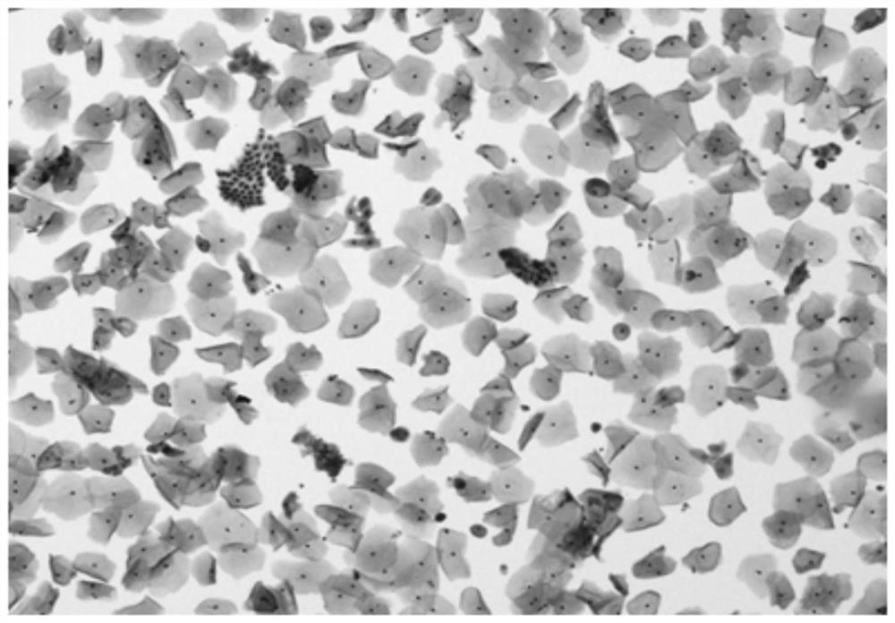
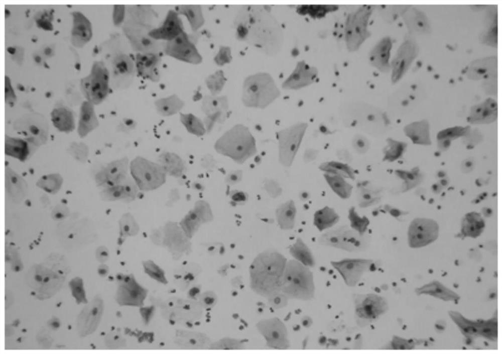
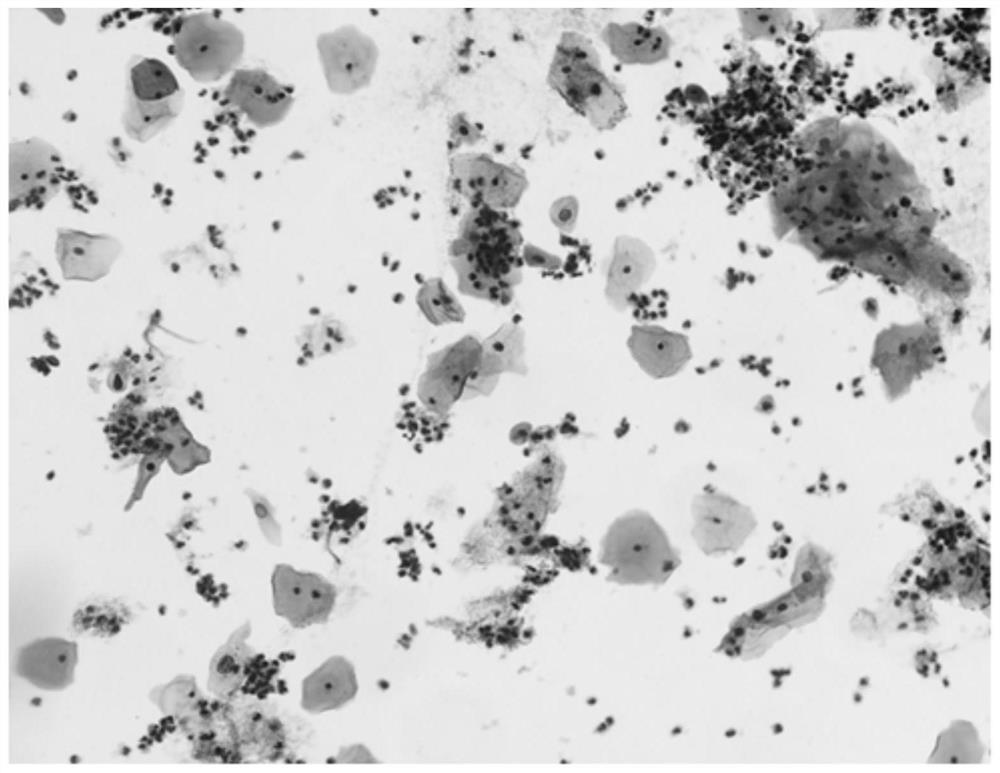
experiment example 1

[0068] Using the cell preservation solution provided in Example 1, the method of Example 4 was used to preserve the vaginal and cervical exfoliated cell specimens, sputum specimens, vaginal and cervical bloody specimens, and needle aspiration cell specimens, and then make slides for microscopic examination to observe the cell morphology. see results Figure 1-Figure 4 .


PUM


Abstract

The invention discloses cell preserving fluid, a preparation method thereof and a cell preserving method, and relates to the technical field of cell preservation. The cell preserving fluid disclosed by the invention contains ethanol, Tris, EDTA.2Na, bromelain, tris(2-carboxyethyl)phosphine hydrochloride, diisobutyl naphthalene sulfonic acid, BSA, trehalose, procline300 and glacial acetic acid. The cells preserved by the cell preserving fluid are complete in morphological structure, interference components such as mucus and red blood cells can be effectively cracked, interference to a detection result is avoided, and a good foundation is provided for subsequent slide preparation and dyeing.

Description

technical field [0001] The invention relates to the technical field of cell preservation, in particular to a cell preservation solution, a preparation method thereof, and a cell preservation method. Background technique [0002] Cytopathological examination is to observe the pathological morphology and make a qualitative diagnosis by exfoliating, scraping and puncturing the cells extracted from the lesion of the patient. It is mainly used in the diagnosis of tumors, and can also be used in the examination and diagnosis of certain diseases. Such as the diagnosis of internal organ inflammatory diseases and the judgment of hormone levels. Cytological specimens can be exfoliated cells from the secretions and excretions of the reproductive tract, respiratory tract, digestive tract, urinary tract, etc., or can be extracted from the chest, abdomen, pericardial cavity, joint cavity, and cerebrospinal fluid through puncture. exfoliated cells, or cells from lesions in various tissues...

Claims


Application Information

Patent Timeline
Patent Type & Authority Applications(China)
IPC IPC(8): A01N1/02
CPCA01N1/021
Inventor 刘飞鸿
Owner 深路医学科技(武汉)有限公司
Why Eureka
Try Eureka