Method for fabrication of three-dimensional lung organoid comprising human stem cell-derived alveolar macrophage

A technology of alveolar macrophages and organoids, applied in artificial cell constructs, biochemical equipment and methods, specific-purpose bioreactors/fermenters, etc., can solve problems such as no lung organoid methods

Pending Publication Date: 2021-05-14
KOREA RES INST OF CHEM TECH
View PDF4 Cites 3 Cited by
  • Summary
  • Abstract
  • Description
  • Claims
  • Application Information

AI Technical Summary

Problems solved by technology

However, there is no known method for producing lung organoids containing mature human alveolar macrophages

Method used


Image

  • Method for fabrication of three-dimensional lung organoid comprising human stem cell-derived alveolar macrophage
  • Method for fabrication of three-dimensional lung organoid comprising human stem cell-derived alveolar macrophage
  • Method for fabrication of three-dimensional lung organoid comprising human stem cell-derived alveolar macrophage

Examples


Embodiment 1

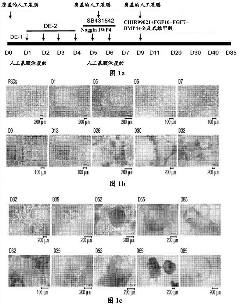
[0090] Example 1: Preparation of three-dimensional lung organoids

[0091] coated board

[0092] The artificial basement membrane matrix that can simulate the extracellular microenvironment (artificial basement membrane-hESC qualified matrix, Cat.#.354277, Corning, USA) was diluted 200 times with cold DMEM / F12 medium (final artificial basement membrane concentration: 250~290μl / 500ml), and put into a 4-well plate (400μl / well), then at 37℃, 5% CO 2 Coat for 1 hour in the incubator.

[0093] Day 1 of differentiation

[0094] Each well of the cell culture plate was coated by adding 10 μg of vitronectin XF (Cat. #.07180, Stem cell technologies) to the plate. By heating at 37°C and 5% CO2 in the presence of TeSR TM -E8 TM Human pluripotent stem cell line H9 (Wicell, WA09-FD) was prepared by culturing in coated plates with culture medium (Cat.#.05940, Stem cell technologies) (2 ml / well). At this time, the medium was changed every day during subculture.

[0095] After r...

experiment example 1

[0120] Experimental example 1: Confirmation of cell morphology in 2D culture and 3D culture after differentiation on day 20

[0121] During the differentiation of hPSCs, the cell morphology was observed by taking pictures while the 2D culture was continued even after the 20th day of differentiation.

[0122] In particular, cells on days 0, 1, 5, 6, 7, 9, and 13 of differentiation, cells on days 26, 30, and 33 of differentiation in which 2D culture was continued, and cells in which differentiation after day 20 were photographed using a microscope Cells at days 32, 35, 52, 65, and 85 of differentiation were changed from two-dimensional culture to three-dimensional culture, and the cell shape was observed.

[0123] As a result, when the cells were differentiated by two-dimensional culture until day 33, tufted cell aggregates were observed, whereas cells differentiated by three-dimensional culture without artificial basement membrane differentiated into clusters similar to actua...

experiment example 2

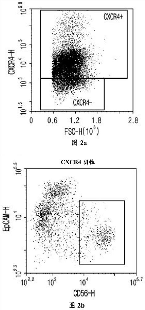
[0124] Experimental example 2: Confirmation of the expression of CXCR4 markers and mesoderm cell markers on the 4th or 5th day of differentiation

[0125] On day 4 or 5 of differentiation, when the proportion of cells expressing CXCR4 (a complete endoderm (DE) marker) reached 70% to 80%, the cell surface was stained for CXCR4, EpCAM, and CD56 and analyzed by flow cytometry. The degree of differentiation was analyzed technically.

[0126] Specifically, on day 4 or 5 of differentiation, remove the medium of the cells, wash the cells with DPBS, and then add TrypLE TM Express, then incubate for 3 to 5 minutes. After that, remove TrypLE TM Express, and cells were collected by adding DPBS, followed by centrifugation at 1,300 rpm for 5 min. After removing DPBS, cells were suspended in FACS buffer. 2 μl of each antibody (CXCR4, cat.#.551510, BD pharmigen; EpCAM, Cat.#.324204, Biolegend; CD56, Cat.#.555518, BD pharmigen) was added to 1×10 4 ~1×10 5 cells, and react at room tem...


PUM


Abstract

The present invention relates to a method for fabrication of a three-dimensional lung organoid comprising human stem cell-derived alveolar macrophages. Specifically, a lung organoid is fabricated by co-culturing cells not expressing the definitive endoderm marker CRCX4 according to a fabrication method of the present disclosure. The lung organoid comprises type 1 and type 2 alveolar epithelial cells as well as alveolar macrophages and realizes infectious or inflammatory responses unlike conventional lung organoids that contain no immune cells and as such, can be advantageously used in studying mechanisms of related lung diseases, excavating biomarkers, developing therapeutic agents, and so on.

Description

technical field [0001] The present invention relates to a method for producing three-dimensional lung organoids comprising human stem cell-derived alveolar macrophages. Background technique [0002] Recently, fine dust has become an important environmental problem that threatens people's safety and health. The pollution level of fine dust is continuously increasing, and its sensory index is also getting higher and higher. Depending on particle size, fine dust with a diameter of 10 μm or less is classified as PM10, fine dust with a diameter of 5 μm or less is classified as PM5, fine dust with a diameter of 2.5 μm or less is classified as PM2.5, and fine dust with a diameter of 1 μm or lower fine dust is classified as PM1. Fine dust with a diameter of 2.5 μm or less is called "ultrafine dust". [0003] According to recent studies, chronic obstructive pulmonary disease (COPD), lung inflammation, emphysema, airway remodeling, etc. have been reported in rat models exposed to fi...

Claims


Application Information

Patent Timeline
Patent Type & Authority Applications(China)
IPC IPC(8): C12N5/071
CPCC12M21/08C12M23/58C12N5/0697C12N5/0645C12N5/0688C12N2533/90C12N2501/15C12N2501/155C12N2501/415C12N2501/117C12N2501/119C12N2501/385C12N2501/727C12N2513/00C12N2501/10C12N2501/999C12N2533/54
Inventor 金银美金基锡李香爱
Owner KOREA RES INST OF CHEM TECH
Why Eureka
Try Eureka