


Quick Research
Generate reliable direction feasibility study reports for your R&D in just a few steps.

Technical Q&A
Discover and master advanced knowledge NOW. Basics, ideas, possibilities, all at once.

Find Solutions
As an expert in R&D theories, this can generate solutions to your technical problems instantly.

Evaluate Feasibility
Analyze your overall solution with one click, know your potential R&D risks in advance.

Monitor Landscape
Get weekly tech updates, stay abreast of the latest tech innovations and key insights.
Application of Tff1 in vascular diseases
A blood vessel and disease technology, applied in the application field of Tff1 in vascular diseases, can solve the problems of complex reasons and no literature reports on trefoil factor 1
- Summary
- Abstract
- Description
- Claims
- Application Information
AI Technical Summary
Problems solved by technology
Method used
Image

Examples
Embodiment 1
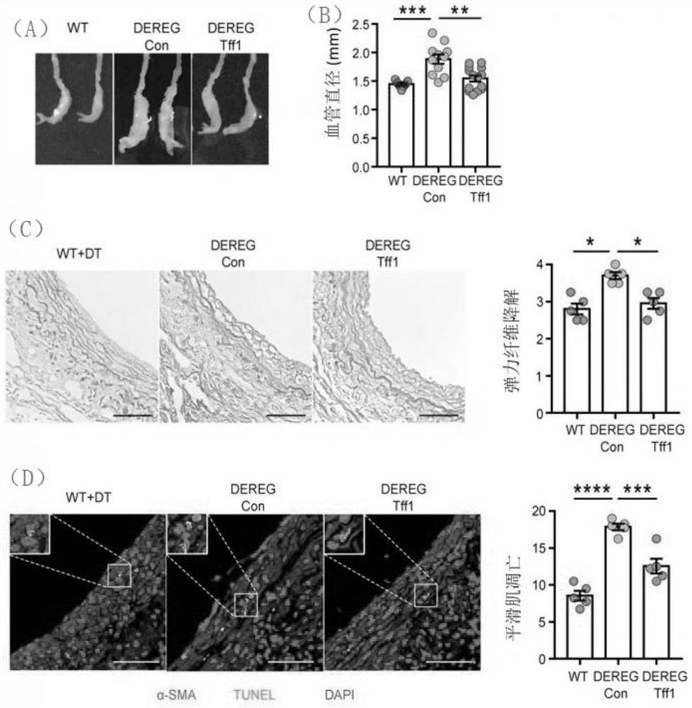
[0037] 1. Establishment of the abdominal aortic aneurysm model induced by elastase
[0038] Male C57BL / 6 or DEREG mice aged 8-12 weeks were anesthetized by intraperitoneal injection of 1% pentobarbital sodium (50mg / kg), fixed on the mouse board with their backs facing down, and the model was established under a stereo microscope : Cut the skin and subcutaneous fascia with ophthalmic scissors from the center of the linea alba in order to make a longitudinal incision about 1.5 cm in length to expose the abdominal viscera, and use a small cotton swab to push the intestinal tube to both sides to fully expose the abdominal aorta and inferior cavity For the vein part, the abdominal aorta was carefully separated with microscopic forceps and suture needles, and the cotton cloth soaked with porcine trypsin was evenly covered on the wall of the abdominal aorta for 40 minutes, then the cotton cloth was discarded, and the abdominal cavity was washed with normal saline for 2 Finally, the i...
Embodiment 2
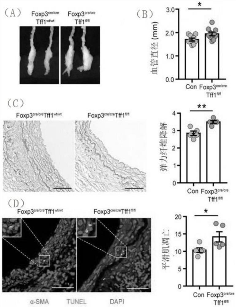
[0053] 1. Establishment of the abdominal aortic aneurysm model induced by elastase
[0054] 8-12 week-old male Foxp3cre / creTff1wt / wt or Foxp3cre / creTff1fl / fl mice were anesthetized by intraperitoneal injection of 1% sodium pentobarbital (50mg / kg), fixed on the mouse board with their backs down, and in vivo Under the microscope, start to build the model: cut the skin and subcutaneous fascia with ophthalmic scissors in sequence from the center of the abdominal linea alba to expose the abdominal viscera. For the abdominal aorta and inferior vena cava, the abdominal aorta was carefully separated with microscopic forceps and suture needles, and the cotton cloth soaked with porcine trypsin was evenly covered on the wall of the abdominal aorta for 40 minutes, and then the cotton cloth was discarded. The abdominal cavity was washed twice with normal saline, and the incision was sutured to close the abdominal cavity.
[0055] 2. Experimental grouping
[0056] (1) Control group: Foxp3...
Embodiment 3
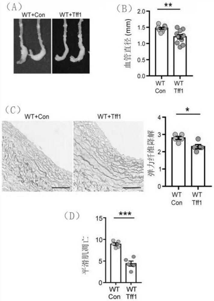
[0068] 1. Establishment of the abdominal aortic aneurysm model induced by elastase
[0069] Male C57BL / 6 mice aged 8-12 weeks were anesthetized by intraperitoneal injection of 1% pentobarbital sodium (50 mg / kg), fixed on the mouse board with their backs facing down, and the model was established under a stereomicroscope: from In the middle of the linea alba, cut the skin and subcutaneous fascia in turn with ophthalmic scissors to make a longitudinal incision about 1.5 cm in length to expose the abdominal viscera, and use a small cotton swab to push the intestinal tube to both sides to fully expose the abdominal aorta and inferior vena cava , carefully separate the abdominal aorta with microscopic forceps and suture needles, and evenly cover the abdominal aortic wall with cotton cloth soaked with porcine trypsin for 40 minutes, then discard the cotton cloth and rinse the abdominal cavity twice with normal saline. Finally, the incision was sutured to close the abdominal cavity. ...
PUM
 Abstract
Description
Claims
Application Information
 - R&D Engineer
- R&D Manager
- IP Professional
- Industry Leading Data Capabilities
- Powerful AI technology
- Patent DNA Extraction
Browse by: Latest US Patents, China's latest patents, Technical Efficacy Thesaurus, Application Domain, Technology Topic, Popular Technical Reports.
© 2024 PatSnap. All rights reserved.Legal|Privacy policy|Modern Slavery Act Transparency Statement|Sitemap|About US| Contact US: help@patsnap.com