Kit for rapidly detecting peripheral T cell lymphoma and use method thereof

A kit and lymphoma technology, applied in the field of immunological detection, can solve the problems of complex steps, large sample volume of the kit, and low detection efficiency, and achieve the effects of reducing the detection cost, shortening the detection period, and reducing double damage

Pending Publication Date: 2021-06-18
河南凯普瑞生物技术有限公司
View PDF0 Cites 2 Cited by
  • Summary
  • Abstract
  • Description
  • Claims
  • Application Information

AI Technical Summary

Problems solved by technology

[0005] In view of the above-mentioned technical problems, the present invention proposes a kit for rapidly detecting peripheral T-cell lymphoma and its use method, which solves the technical problems of large sample size, complicated steps, and low detection efficiency required by existing kits

Method used


Image

  • Kit for rapidly detecting peripheral T cell lymphoma and use method thereof
  • Kit for rapidly detecting peripheral T cell lymphoma and use method thereof
  • Kit for rapidly detecting peripheral T cell lymphoma and use method thereof

Examples


Embodiment 1

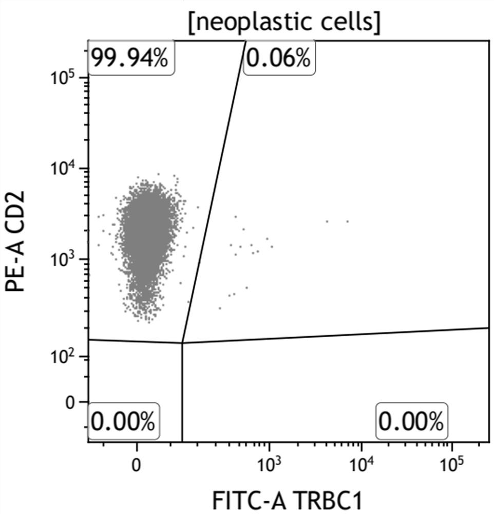
[0039] Utilize the test kit of the present application, to a case that has definite clinical diagnosis such as figure 1 clinical samples for testing. In this example, a flow cytometer is used. The operation steps of the test are as follows:

[0040] Add hemolysin with a volume equivalent of about 1:20 to 100 μL sample and incubate at room temperature in the dark for 10 minutes to remove cell debris or non-cellular components. Repeat the operation to prepare two samples. After adding a small amount of sheath fluid to dilute, mix two groups of monoclonal antibody mixtures Add 20 μL to the two samples one by one at room temperature and incubate in the dark for 15 minutes, add 500 μL of PBS solution and incubate in the dark for 5 minutes at room temperature, centrifuge and then dilute with an appropriate amount of sheath solution to be tested on the machine.

[0041] The test data obtained from the experiment, such as figure 1 shown. Tumor T cells express monotype TCRβ, and TR...

Embodiment 2

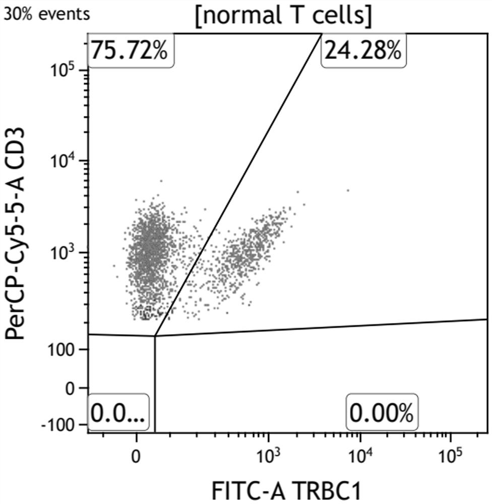
[0043] Using the kit of this application to detect a normal clinical sample, the flow cytometer used, the specific operation steps are: take 100 μL samples respectively, add hemolysin with a volume equivalent of about 1:20, and incubate at room temperature for 15 minutes in the dark Centrifuge twice to remove cell debris and non-cellular components, then add a small amount of sheath fluid, add 20 μL of the two monoclonal antibody mixtures one by one, incubate at room temperature for 15 minutes, add 2 mL of sheath fluid and centrifuge again, and then add 500 μL of sheath fluid The machine performs two tests.

[0044] The final test results are as figure 2 As shown, the TRBC1 antibody test results showed partial expression in normal samples.

[0045] The results show that the TRBC1 antibody kit used in this patent can realize the sensitive diagnosis of PTCL patients, and simply and accurately screen T cell subsets and reactive lymphocytes whether TCRβ presents monotypic expres...

Embodiment 3

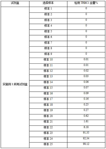
[0047] Using the kit of the present application, 25 cases of mature T-cell lymphoma samples invading bone marrow were detected simultaneously.

[0048] In this embodiment, a flow cytometer is used, and the operation steps of each test are as follows:

[0049] Take 20 μL of monoclonal antibody mixture and 100 μL sample and incubate at room temperature in the dark for 15 minutes, add hemolysin at a volume equivalent of about 1:20 and incubate at room temperature in the dark for 10 minutes, add 500 μL of PBS solution and incubate for 5 minutes at room temperature in the dark, and then test on the machine . Repeat the above steps to obtain 25 sets of detection data;

[0050] As shown in table 2:

[0051] 25 groups of data that table 2 embodiment 3 obtains

[0052]

[0053] It is generally considered that less than 15% or more than 85% of cells with TRBC1 expression are monomorphic in expression of TCRβ. The final results of the test on 25 samples showed that the expression ...


PUM


Abstract

The invention belongs to the field of immunological detection, and relates to a kit, in particular to a kit for rapidly detecting peripheral T cell lymphoma and a use method of the kit. The kit comprises a reagent I and a reagent II, the reagent I comprises two groups of monoclonal antibody mixtures, and the first group of monoclonal antibody mixture comprises TRBC1, CD2, CD3, CD5, CD7, CD16, CD56, CD57 and CD45; the second group of monoclonal antibody mixture comprises the following components: TRBC1, TCR-gamma delta, CD3, CD8, CD4, CD7 and CD45; and the reagent II is hemolysin. The kit provided by the invention realizes the rapid and accurate detection of the peripheral T cell lymphoma (PTCL), is simple and convenient, greatly reduces the requirement on the sample size, and reduces the burden of a patient.

Description

technical field [0001] The invention belongs to the field of immunological detection and relates to a kit, in particular to a kit for rapidly detecting peripheral T-cell lymphoma and a use method thereof. Background technique [0002] Currently, the immunophenotypic diagnosis of T-cell lymphoma depends on the loss or abnormality of T-cell surface antigens. However, there are many types of T-cell lymphoma, the classification is complicated, and pathological diagnosis is difficult. These changes are often very weak, and the diagnosis of some T-cell lymphomas is difficult to clarify the nature only by HE and immunohistochemical staining; while flow cytometry can detect the absence, abnormal increase or decrease of T-cell antigen expression to assist T-cell lymphoma. T cell lymphoma diagnosis, but some T cell lymphomas have no obvious phenotypic abnormalities or activated reactive T cells during infection and T cells may also have phenotypic abnormalities in autoimmune diseases,...

Claims


Application Information

Patent Timeline
Patent Type & Authority Applications(China)
IPC IPC(8): G01N33/577G01N33/574G01N33/569
CPCG01N33/577G01N33/57407G01N33/57492G01N33/56966
Inventor 王辉岳保红王慧君刘玉珂马军
Owner 河南凯普瑞生物技术有限公司
Why Eureka
Try Eureka