Method and kit for evaluating biological activity of LAG3 antibody
A biological activity and kit technology, applied in the field of LAG3 antibody activity detection, can solve the problems of cumbersome process, poor experimental stability, long construction period, etc., and achieve the effect of short construction time, good stability and effective detection of activity.
- Summary
- Abstract
- Description
- Claims
- Application Information
AI Technical Summary
Problems solved by technology
Method used
Image

Examples
Embodiment 1
[0041] Construct a stable expression of people lag3 Jurkat stabilized cell line.
[0042] Jurkat cells did not express endogenous human LAG3 protein (HLAG3), and the Jurkat stabilized cell line with stable expression of HLAG3 protein (sequence such as SEQ ID NO.1) was constructed by a slow viral system, which is Jurkat-HLAG3, which is specifically as follows.
[0043] 293T cells were seeded in a 100 mm plate using a 10% DMEM complete medium in 5 × 10E6, at 37 ° C, 5% CO 2The incubator was cultured overnight. The next day, to be 293T cells grow to 80 ~ 90%, using polyethylene imine PEI, plasmid transfection, 37 ° C, 5% CO 2 Static culture in the incubator.
[0044] On the second day, the supernatant was completely sucked (the first virus supernatant), and the fresh DMEM complete medium was added and cultured in the incubator for 48 hours. After absorbing the virus, the addition of fresh fully medium continued to cultivate for 1 day, collect the superprint, and combined the supernat...
Embodiment 2
[0048] Evaluation of the Evaluation of the Birage of JURKAT-HLAG3 Stabilized Cell Lines.
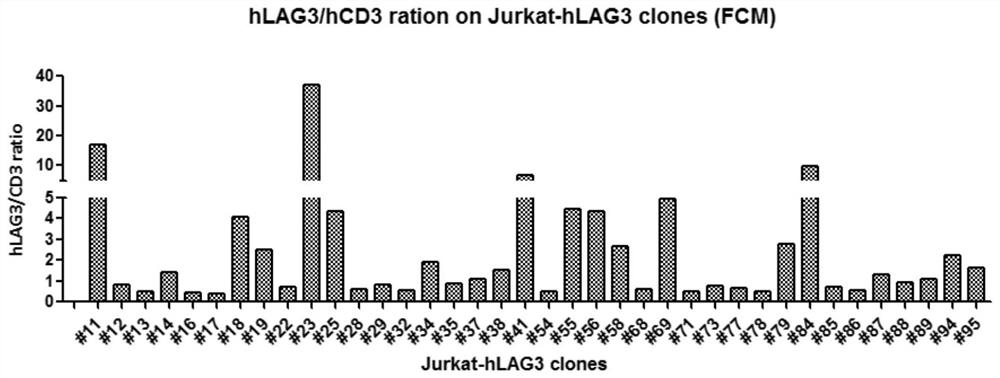
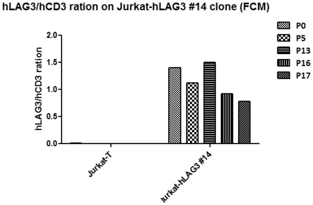
[0049] In order to prevent the loss of the exogenous LAG3 gene, the part Jurkat-HLAG3 clone obtained in Example 1 was stabilized.
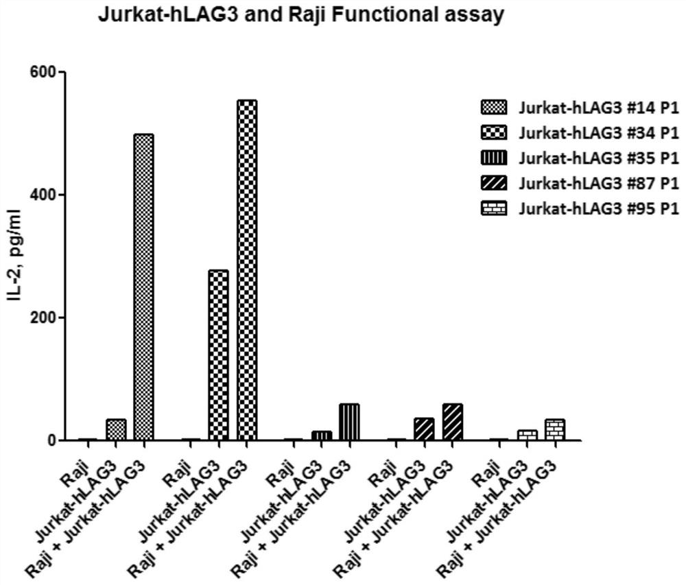
[0050] The secretion of cytokine IL2 is detected by Jurkat-HLAG3 and RAJI cellular mixed culture systems:
[0051] The 5 μg / ml CD3 antibody package was incubated in 96-well plates, the next day, according to 1 × 10E4 / hole Raji cell, 2 × 10E5 / hole Jurkat-HLAG3 cells, 37 ° C continuously for 3 days, enzyme-linked immunization Reaction ELISA detection expression of IL2 in supernatants, test results figure 2 Indicated.
[0052] by figure 2 It can be known, relative # 34, clonal, # 14, clone, clone jurkat-hlag3 almost no IL2 secretion, after adding Raji cells, the secretion of IL2 is significantly up-regulated, indicating that # 14 cells can be obvious Evaluation of biological activity of LAG3 antibodies. At the same time, through flow cytometry, the expression...
Embodiment 3
[0054] A method of evaluating the biological activity of LAG3 antibody.
[0055] The RPMI 1640 fully medium retained RAJI cells containing 10% FBS was 5 × 10E6 / ml of the live cell density of 10% FBS, and the mixed methacin C was added to the Raji cells to the final concentration of 2.5 μg / mL, 37 ° C cell culture. The box is standing for 30 min. After adding DPBS to 10ml, 250 x g from centrifugation 5 minutes, after discarding the supernatant, the cells were washed again with DPBS once.
[0056] Finally, the cells complete medium retained Raji cells to a live cell concentration of 2 × 10E5 / mL. JURKAT-HLAG3 # 14 (Example 2) The cell fluid, 250 × g centrifugation was centrifuged for 5 min, and the live cell concentration to 1 × 10E6 / ml were adjusted.
[0057] After mixing Raji cells and Jurkat-HLAG3 # 14 cells, followed by 100 μl / pore to the 96-well flat bottom plate. Dilution anti-HLAG3 antibodies were used in cells to the desired concentration. Press 100 μL / hole into a ...
PUM
 Abstract
Description
Claims
Application Information
 - R&D Engineer
- R&D Manager
- IP Professional
- Industry Leading Data Capabilities
- Powerful AI technology
- Patent DNA Extraction
Browse by: Latest US Patents, China's latest patents, Technical Efficacy Thesaurus, Application Domain, Technology Topic.
© 2024 PatSnap. All rights reserved.Legal|Privacy policy|Modern Slavery Act Transparency Statement|Sitemap