Quantitative detection method based on ophthalmic retina OCT image

A quantitative detection method and retinal technology, applied in image enhancement, image analysis, image data processing, etc., to improve the treatment effect, save medical resources, and increase the detection rate

Pending Publication Date: 2021-07-02
EYE & ENT HOSPITAL SHANGHAI MEDICAL SCHOOL FUDAN UNIV
View PDF1 Cites 0 Cited by
  • Summary
  • Abstract
  • Description
  • Claims
  • Application Information

AI Technical Summary

Problems solved by technology

It realizes fast and accurate identification of different types of DME, but the technical solution it adopts is based on deep learning, and obtains the results of binary classification tasks with binary classification functions and preset thresholds, so as to realize fast and accurate identification of different types of DME. Accurate identification, which cannot be used for the identification of non-structural lesions in the present invention

Method used


Image

  • Quantitative detection method based on ophthalmic retina OCT image
  • Quantitative detection method based on ophthalmic retina OCT image
  • Quantitative detection method based on ophthalmic retina OCT image

Examples


Embodiment 1

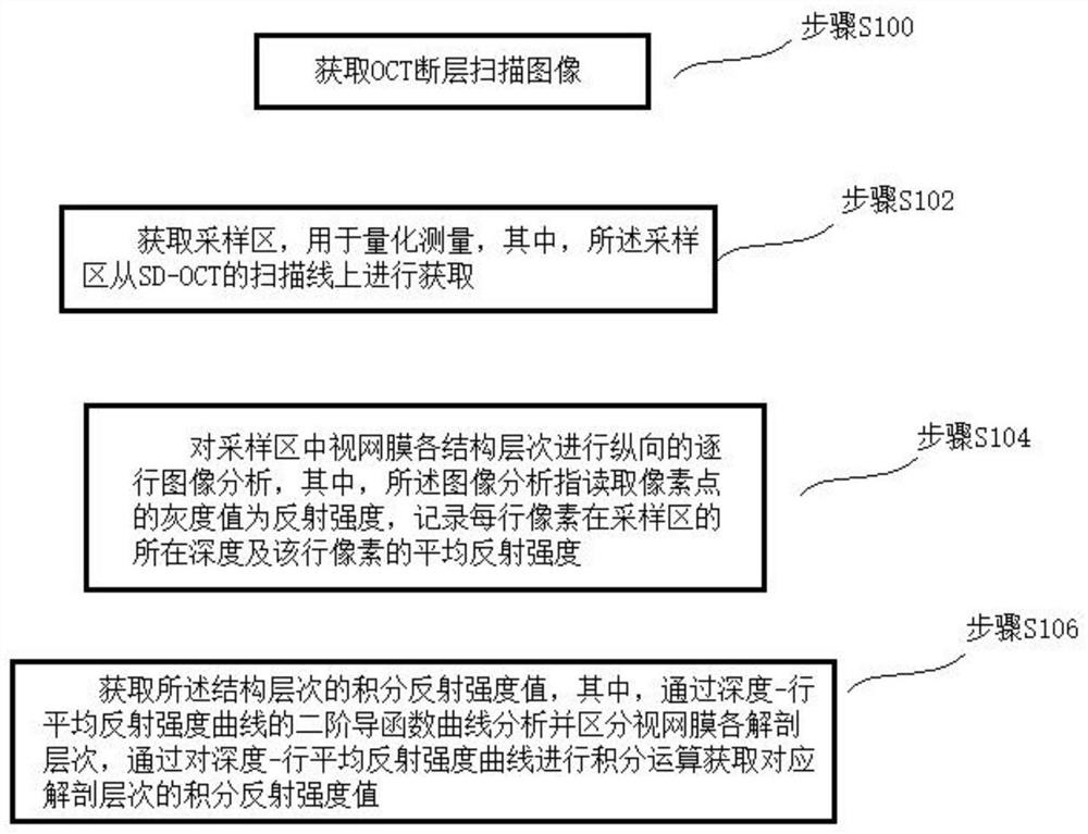
[0022] Please see attached figure 1 , with figure 1 It is a flowchart of a quantitative detection method based on ophthalmic retinal OCT images of the present invention. A quantitative detection method based on ophthalmic retinal OCT images, at least comprising the following steps:

[0023] Step S100: acquiring an OCT tomographic image;

[0024] In some preferred embodiments, SD-OCT is used for scanning, the scanning range is 30°×30°, centered on the fovea of ​​the macula, single-line scanning, multi-line scanning or radiographic scanning is performed, and the automatic real-time average function takes ART=100 frames to obtain retinal OCT images; the scanning mode can be selected but not limited to the above parameters, and the actual parameters can be determined according to the lesion to be detected;

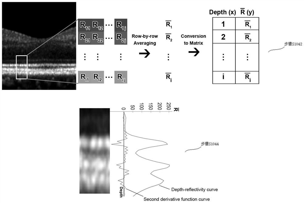
[0025] Step S102: Obtain a sampling area for quantitative measurement, wherein the sampling area is acquired from the scanning line of SD-OCT;

[0026] In some preferred e...

Embodiment 2

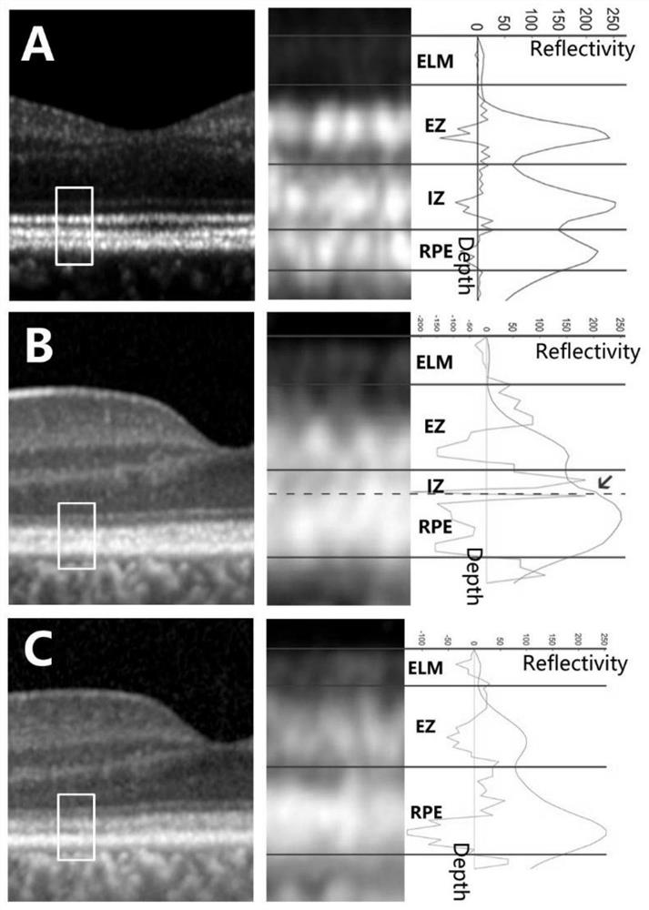
[0044] This embodiment explains the present invention through corresponding application:

[0045] Select the normal control group: select healthy volunteers, inclusion criteria: best corrected visual acuity ≥ 0.8, spherical equivalent refractive power between (inclusive) +1D to -3D; exclusion criteria: ophthalmic surgery history, trauma history , posterior staphyloma, and other history of ophthalmic disease affecting the outer retina.

[0046] Selected disease group: select patients with fundus lesions involving the outer retina, including: non-infectious inflammatory retinopathy (including white dot syndrome, tumor-related retinopathy, uveitis, retinal vasculitis), infectious uveitis ( Including tuberculous uveitis, syphilitic uveitis, ocular toxoplasmosis), macular hole surgery, retinal detachment surgery (including rhegmatogenous retinal detachment, exudative retinal detachment), retinal dystrophy disease (including Retinitis pigmentosa, cone-rod dystrophy, cone dystrophy,...


PUM


Abstract

The invention relates to a quantitative detection method based on an ophthalmology retina OCT image. The quantitative detection method comprises the following steps: acquiring an OCT tomography image; sampling points are obtained and used for quantitative measurement, and the sampling points are located on a scanning line of SD-OCT; carrying out image analysis on a plurality of longitudinal structure layers on the sampling points, wherein the image analysis refers to analysis of gray values of pixel points; obtaining an integral reflection intensity value of the structural hierarchy, obtaining a pixel depth i and a corresponding row average gray value, and constructing a depth-row average reflection intensity curve and a second derivative function curve thereof by taking i as an X axis and a Y axis to obtain integral reflection intensity. The method has the advantages that in some early retinal cases with normal retinal structures, the change of the spatial cumulative reflection intensity of a specific affected structure can be quantitatively reflected, so that the discovery probability of early latent retinal diseases is improved.

Description

technical field [0001] The invention relates to the field of optical coherence tomography data processing, in particular to a quantitative detection method based on ophthalmic retinal OCT images. Background technique [0002] Spectrum-domain optical coherence tomography (SD-OCT) is an auxiliary inspection technique widely used in the field of ophthalmology, especially in retinal diseases. value. [0003] The retinal tomographic images obtained by SD-OCT mainly rely on manual interpretation by clinicians, and have a high detection and recognition rate for diseases with abnormal retinal structures (such as retinal detachment, macular hole, macular epiretinal membrane, etc.). However, for non-structural lesions, especially early outer retinal lesions (such as early retinitis pigmentosa, occult macular degeneration, macular dystrophy, etc.), SD-OCT images often have no obvious structural changes, only showing some The abnormality of some specific levels of reflectivity is diff...

Claims


Application Information

Patent Timeline
Patent Type & Authority Applications(China)
IPC IPC(8): G06T7/00A61B3/00A61B3/10A61B3/12
CPCA61B3/0025A61B3/102A61B3/1225G06T7/0012G06T2207/10101G06T2207/30041
Inventor 常青顾骏祥蒋婷婷俞铭蓉李文婷刘师学陈文文张娟
Owner EYE & ENT HOSPITAL SHANGHAI MEDICAL SCHOOL FUDAN UNIV
Why Eureka
Try Eureka