Tiny residual focus detection method and device, storage medium and equipment

A detection method and residual technology, applied in proteomics, medical data mining, instruments, etc., can solve the difficulties of judging the authenticity, unsatisfactory accuracy of small lesions, lack of correction methods, etc., to improve the credibility , Eliminate noise signals, and accurately detect the effect

Active Publication Date: 2021-07-09
臻和(北京)生物科技有限公司 +2
View PDF8 Cites 3 Cited by
  • Summary
  • Abstract
  • Description
  • Claims
  • Application Information

AI Technical Summary

Problems solved by technology

ctDNA is a molecular marker of minimal residual disease (Minimal Residual Disease) in patients. The detection of minimal residual disease needs to identify a very small amount of ctDNA signal in blood. The difficulty lies in how to improve the possibility of obtaining ctDNA signal and determine the authenticity of low-frequency ctDNA signal. In the existing technology, on the one hand, in order to detect rare ctDNA signals more sensitively, avoiding missed detection is often achieved by expanding the detection range and tracking more variant signals, and multivariate tracking brings new specificity Currently, there is a lack of effective correction methods; on the other hand, there is a lack of effective means to distinguish low-frequency ctDNA signals from NGS platform noise signals, and it has always been a difficult point to judge the authenticity of ctDNA signals; The accuracy of the detection of small lesions is not ideal

Method used


Image

  • Tiny residual focus detection method and device, storage medium and equipment
  • Tiny residual focus detection method and device, storage medium and equipment
  • Tiny residual focus detection method and device, storage medium and equipment

Examples


Embodiment 1

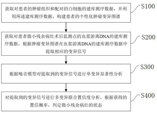
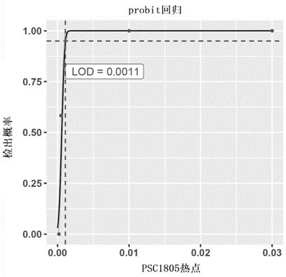
[0145] Example 1: Performance analysis of hotspot-driven single mutation detection based on method 1

[0146]This example analyzes the experimental data for performance verification, based on the sensitivity and specificity of method 1 for hotspot-driven single variant detection. In this analysis performance verification experiment, the UMI (Unique Molecular identifier, UMI) molecular tag adapter was used to construct the library, and then the PanelP1 (Table 1.1) was used to enrich the target region. PanelP1 covered the 108Kb interval of 29 genes , the enriched library was subjected to high-depth sequencing. In the sensitivity assessment, 12 known hotspot-driven variations were used to make a standard positive sensitivity control-PSC1805 (see Table 1.2); in the specificity assessment, cfDNA from 149 healthy individuals was used to evaluate 19 Specificity of detection of driver variants in tumor hotspots.

[0147] 1.1 Sensitivity and minimum detection limit based on method 1 ...

Embodiment 2

[0169] Example 2: Performance Analysis of Single Variation Detection Based on Methods 1, 2, and 3

[0170] In this example, by analyzing the experimental data of performance verification, the sensitivity and specificity of the three analysis processes for the detection of non-hotspot single mutations are verified based on three different methods. The KAPA Hyper Preparation Kit (Roche Diagnostics product) was used to construct the library, and then the PanelP2 (see Table 2.1) was used to enrich the target region. PanelP2 covered the 2.1Mb interval of 769 genes. The enriched library was highly deep sequencing. During the performance evaluation, the samples used were prepared by mixing the white blood cell DNA of a single individual S with known SNP site information and the negative control negative standard GM12878.

[0171] 2.1 Sample information

[0172] The 32 SNP variations (single nucleotide mutations) of individual S different from hg19 (human genome version) and GM12878...

Embodiment 3

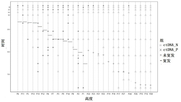
[0191] Example 3: Analysis of sample detection performance during multivariate tracking (based on method 1)

[0192] Since the content of cfDNA in blood limits the detection sensitivity of single mutations, method 1 can significantly improve the overall detection sensitivity by simultaneously tracking prior tumor-specific mutations in multiple tissues. In the MAVC2006 series of samples, different ratios of mixed DNA were used to simulate plasma DNA with different tumor proportions. In order to reduce the influence of site sampling, 100 random samplings were carried out by computer for each specified number, that is, 100 independent tumor prior variation maps were formed. For a diluted sample, each time according to each group The map carries out the mutation signal tracking of the designated site and determines the status of the small residual lesions. A total of 100 determinations are required. Finally, the positive detection rate of the 100 samples was counted as the detect...


PUM


Abstract

The invention discloses a tiny residual focus detection method and device, a storage medium and equipment, and belongs to the technical field of biological detection. The method comprises the following steps of: acquiring library building and sequencing data of tumor tissues and paired white blood cells of a patient, and constructing a personalized tumor variation map of the patient by utilizing the library building and sequencing data; acquiring library building and sequencing data of plasma free DNA of the postoperative monitoring point of the tiny residual focus of the patient, and extracting a corresponding variation signal from the library building and sequencing data of the plasma free DNA according to the tumor variation map; performing single variation significance analysis on the extracted variation signal according to a noise model which is a combined model; and carrying out multi-variation joint confidence analysis on the extracted variation signal, and judging the state of the tiny residual focus according to the obtained confidence probability. The invention further provides the corresponding device, the storage medium and equipment. According to the tiny residual focus detection method and device, the storage medium and the equipment of the invention, tiny residual focuses can be accurately detected.

Description

technical field [0001] The invention belongs to the technical field of biological detection, and in particular relates to a detection method, device, storage medium and equipment for tiny residual lesions. Background technique [0002] Regular detection of minimal residual lesions can provide effective reference for doctors to choose tumor treatment methods, treatment cycles, medication guidance, and patient drug resistance tracking. ctDNA is a molecular marker of minimal residual disease (Minimal Residual Disease) in patients. The detection of minimal residual disease needs to identify a very small amount of ctDNA signal in blood. The difficulty lies in how to improve the possibility of obtaining ctDNA signal and determine the authenticity of low-frequency ctDNA signal. In the existing technology, on the one hand, in order to detect rare ctDNA signals more sensitively, avoiding missed detection is often achieved by expanding the detection range and tracking more variant sig...

Claims


Application Information

Patent Timeline
IPC IPC(8): G16B20/20G16B20/50G16B40/10G16H50/20G16H50/70
CPCG16B20/20G16B20/50G16B40/10G16H50/20G16H50/70G16H70/60G16H10/40G16H50/30G16H50/50C12Q1/6886C12Q2600/156G16B40/00C12Q1/6809C12Q1/6874C12Q1/6806C12Q1/6837C12Q2600/158
Inventor 张亚晰谢泓禹陈维之杨滢范锐赵秀玉杨飘于佳宁何骥杜波
Owner 臻和(北京)生物科技有限公司
Why Eureka
Try Eureka