3D printing cerebral hemorrhage model for puncture training and efficient manufacturing method thereof

A 3D printing and model technology, applied in the field of medical teaching, can solve the problems of time-consuming, easy to change, difficult to operate, etc., and achieve the effect of ensuring absolute accuracy, preventing material waste, and accurate skull position

Pending Publication Date: 2021-07-23
SUN YAT SEN UNIV CANCER CENT +1
View PDF0 Cites 3 Cited by
  • Summary
  • Abstract
  • Description
  • Claims
  • Application Information

AI Technical Summary

Problems solved by technology

[0005] 1. Autologous blood injection method: Create a cerebral hemorrhage model by injecting autologous arterial blood or blood clots; although it can simulate the clinical cerebral hemorrhage process, it is still very different from the clinical symptoms of actual patients, and the shape and size of the hematoma are difficult to guarantee , and the repeatability is poor, it is difficult to use for operation training
[0006] 2. Collagenase method: Dissolving small blood vessels by injecting bacterial collagenase into the brain, although it can simulate the process of hematoma expansion and rebleeding, it is a symptom of multiple blood vessels (arteries, veins, capillaries), which is different from A common arterial hemorrhage clinically; and the bleeding method is hemorrhage, lack of acute mass effect, and the symptoms of real cerebral hemorrhage are also very different
However, the position accuracy of the hematoma produced by this method is not high, and it is difficult to be used for accurate measurement and positioning before puncture operation
[0008] 4. Spontaneous cerebral hemorrhage model: There are problems such as difficulty in obtaining breeders, easy mutation, complicated process, and long time-consuming, and the operation is difficult, and the bleeding location and bleeding volume cannot be controlled
To a certain extent, this model can help to be proficient in the process of endoscopic hematoma removal and the use of related instruments. However, due to the large difference between the shape of the model and the clinical cerebral hemorrhage, and the lack of important craniofacial anatomical landmarks, it is impossible to realize the hematoma. Operation exercises related to minimally invasive drainage
[0012] 2. Standardized models: The existing cerebral hemorrhage models on the market are non-individualized cerebral hemorrhage models produced in batches. The properties of their materials are far from those of real clinical patients, and they still remain at the level of observation and learning, and cannot be operated manually the goal of
[0013] To sum up, there is a lack of a cerebral hemorrhage model in the prior art that can completely imitate the pathophysiological changes of cerebral hemorrhage, visually inspect the training effect, low cost, and be used for operation training to accumulate experience through practice

Method used


Image

  • 3D printing cerebral hemorrhage model for puncture training and efficient manufacturing method thereof
  • 3D printing cerebral hemorrhage model for puncture training and efficient manufacturing method thereof
  • 3D printing cerebral hemorrhage model for puncture training and efficient manufacturing method thereof

Examples


Embodiment 2

[0094] Embodiment two: if Figure 11 As shown, on the basis of Embodiment 1, the simulated hematoma 5 is directly positioned at the position of the foramen magnum through the positioning rod 52, and the inside of the hematoma cyst wall 511 can be perfused and filled with the first transparent flexible material directly from the position of the foramen magnum to form a simulated The blood clot 512 does not need to be additionally provided with a skull base base 4, and has a simple structure and is easy to operate.

[0095] When filling the simulated brain tissue 6 , the external auxiliary structure can be used to assist the limit positioning rod 52 and the simulated blood mass 5 .

Embodiment 3

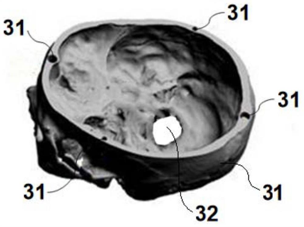
[0096] Embodiment 3: On the basis of Embodiment 1, a plurality of positioning rods are arranged inside the lower skull 3, and a plurality of trays 53 are fixedly supported by the plurality of positioning rods, and a simulated hematoma 51 can be fixedly arranged above each tray 53, To simulate multiple cerebral hematomas; in this embodiment, a maximum of 12 trays can be fixedly set, which can simulate the simultaneous occurrence of 12 hematomas.

[0097] The actual operation process of the present invention is as follows:

[0098] 1. Collect medical digital imaging and communication DICOM (hereinafter referred to as DICOM) data of brain CT of patients with cerebral hemorrhage clinically, import it into a computer with Mimics software installed, open the DICOM data, perform 3D reconstruction, and perform corresponding processing according to the model requirements:

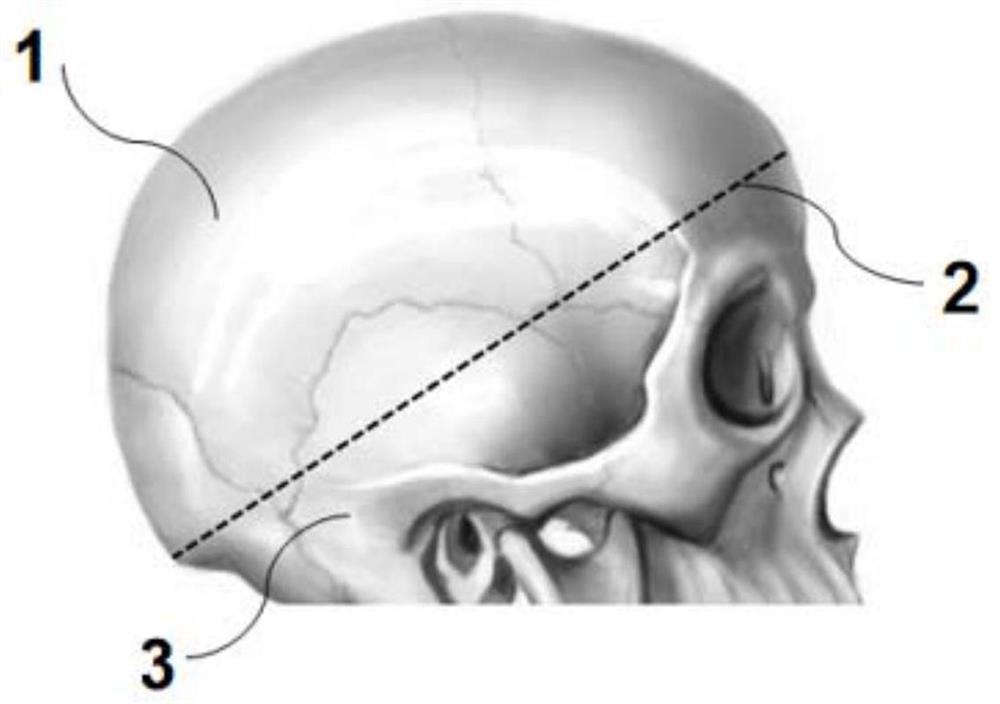
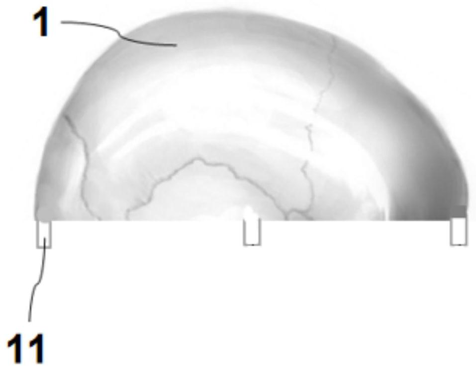
[0099] 1) Skull reconstruction and design processing: After reconstructing the skull with Mimics software, design...


PUM


Abstract

According to the 3D printing cerebral hemorrhage model for puncture training and the efficient manufacturing method of the 3D printing cerebral hemorrhage model, the smooth interface of an upper skull and a lower skull is limited and combined through a limiting groove and a limiting boss to form the whole simulation skull, a flexible simulated hematoma block is installed in the lower skull through a skull seat, and the simulation skull is further filled with a flexible simulated brain tissue; the simulated brain tissue wraps the simulated hematoma block, and the simulated brain tissue and the simulated hematoma block are made of transparent materials; after a 3D model is created by imitating any real cerebral hemorrhage case clinically, an individualized model is manufactured through a 3D printing technology and is used for practical puncture operation training; pathophysiological changes of cerebral hemorrhage can be completely simulated, and puncture experience of the model is vivid; after the operation is completed, the path deformation of the puncture needle can be visually inspected through the transparent simulated brain tissue and the simulated hematoma block, and the puncture effect can be clearly judged; and the device is accurate in position, can be repeatedly used or recycled, and is simple in structure, low in cost and convenient to operate.

Description

technical field [0001] The invention belongs to surgical technique training in the technical field of medical teaching, and in particular relates to a 3D printed cerebral hemorrhage model for puncture training and an efficient manufacturing method thereof. Background technique [0002] Cerebral hemorrhage is a bleeding disorder caused by rupture of blood vessels in the brain parenchyma caused by various etiologies. Its early mortality rate is very high. Most of the survivors have complications and sequelae of varying degrees. Minimally invasive puncture and drainage are used to treat intracranial hematoma. The method is developing rapidly and has the advantages of less trauma, less sequelae and complications, but its surgical effect still depends on the skill level of the operator, and the technical level of the operator needs to be improved through the accumulation of practical experience. The cerebral hemorrhage model in the prior art The production methods are generally d...

Claims


Application Information

Patent Timeline
Patent Type & Authority Applications(China)
IPC IPC(8): G09B23/28B33Y10/00B33Y50/00B33Y80/00
CPCB33Y10/00B33Y50/00B33Y80/00G09B23/28
Inventor 牟永告魏大年余成伟
Owner SUN YAT SEN UNIV CANCER CENT
Why Eureka
Try Eureka